Short Communication - Insights in Nutrition and Metabolism (2018) Volume 2, Issue 3
Hyperhomocysteinemia effects on histology and lipid content of aorta in male and female rabbit.
Othmani-Mecif K*, Fernane A, Taghlit A, Yefsah A, Ghoul A, Benazzoug Y
Laboratory of Cellular and Molecular Biology, Team of ECM Remodeling Biochemistry, Faculty of Biological Sciences, University of Sciences and Technology Houari Boumediene, Algiers, Algeria
- *Corresponding Author:
- Khira Othmani-Mecif
Universite des Sciences et de la Technologie Houari Boumediene Algiers Algeria
Tel: +213 21 24 79 50
E-mail: kothmani@ymail.com
Received date: March 14, 2016; Accepted date: April 19, 2016; Published date: April 22, 2016
Citation: Othmani K, Fernane A, Taghlit A, et al. Hyperhomocysteinemia effects on histology and lipid content of aorta in male and female rabbit. Insights Nutr Metabol. 2019;2(1):5-10.
Abstract
Background: The aim of the study is to compare the effects of hyperhomocysteinemia induced by methionine overload on lipid parameters and structure of the aorta in both male and female rabbits. Materials and methods: Study is carried on 20 male rabbits (control, n=11 and experimented, n=9) and 16 female rabbits, control (n=8) and tested (n=8). Control models are fed standard diet, the Met females and males received standard diet supplemented with Met (500 mg/day) during 3 months. Results obtained from MetM and MetF are compared with those of the respective controls. The body weight is measured, the parameters, Hcy, total cholesterol, total lipids, TG, atherogenicity index ((VLDL+LDL)/ HDL) are determinate by appropriate methods (FPIA, colorimetry). In aorta tissue we assess the total lipids, TC and TG contents and realize a histological study (Masson's staining). Results and discussion: the Met-enriched diet causes a low increase of Hcy (p<0.05) in male, but a significant increase in female (p<0.0001). The TG raise in male (p<0.01) and diminish in female (p<0.01). Atherogenicity index augments from 1,31 to 1,52 in male and from 2,17 to 3,07 in female. At the aortic level, while in male rabbit the total lipid, TC and TG raise significantly (p<0.01), in female TL decrease but TC and TG increase (p<0.01). Aorta of Met female undergoes severe histological changes, such as endothelium hypertrophy, local rupture of the internal elastic lamina, remodelling of media with accumulation of collagens. The observations made in Met male are less important, but the elastic blades present irregular assemblies. Conclusion: Under Met overload, the female aorta tissue presents the most spectacular changes of biochemical as well as histological parameters.
Keywords
Methionine, Hcy, Aorta, Lipid, Male, Female.
Introduction
Met enriched diet causes hyperhomocysteinemia (Hhcy), which is associated with the metabolic syndrome, oxidative and nitrosative endoplasmic reticulum stress [1], inflammation, and increased cardiovascular risk [2]. Hhcy induces HDL changes through Hcy-thiolactone, with loss of their anti-inflammatory and cyto-protective properties [3], and promotes LDL oxidation and internalization by macrophages, the initial step of atherosclerosis [4]. The Hcy autoxidation causes endothelial dysfunction, considered as the main initiator of atherogenesis, via H2O2 [5] reducing endothelial relaxation by fat accumulation. The impact of the diet on the cardiovascular system related to the gender has been studied in some studies, so Nematbakhsh et al. [6] indicate a gender difference in endothelial permeability of aorta in rabbits consuming normal or high-cholesterol diets. For Pektaş et al. [7], the protection with females to metabolic abnormalities could be attributed to estrogen and the type of diet may differently affect metabolic parameters between males and females. Another study, realized in patients with a bicuspid aortic valve indicate more enhanced collagen degradation and smooth muscle cell loss in male than in female patients [8].
Materials and Methods
Study is carried on 20 male rabbits divided into Control (n=11) and experimented (n=9) and 16 female rabbits divided into control (n=8) and tested (n=8). Control models are fed standard diet, the Met females and males received standard diet supplemented with methionine (500 mg/day) during 3 months. All experimental procedures were authorized by the Institutional Animal Care Committee of the National Administration of Algerian Higher Education and Scientific Research, ethical approval number: 98-11, law of 22 august 1998. Results obtained from experimented rabbits are compared with those of the control. The monitoring concerns the body weight measure, the homocystein determination (method FPIA) [9] and the cholesterol [10], triglycerids [11] (Kits Randox) and atherogenicity index ((VLDL+ LDL)/HDL) [12] in the serum. At the end of experiment, we assess the content of the total lipids, TC and TG in aorta lysate. The histological study of the aortic tissues harvested from control and experimented models are stained by Masson’s trichrome, Periodic Acid –Schiff and silver nitrate methods.
Statistical analysis
All data are expressed as mean ± standard error of the mean (SEM). Statistical analyses were achieved by Student’s t test using SPSS 20.0 (SPSS Inc., Chicago, IL). Differences were considered significant at p<0.05.
Results and Discussion
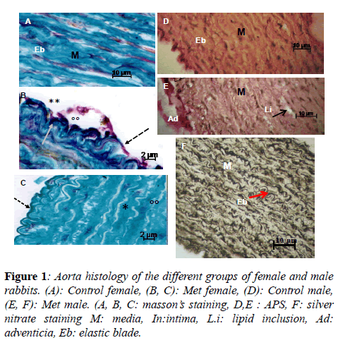
At the end of experimentation, the Met overload causes a low increase of Hcy (p<0.05) in male [13], while in female this augmentation is very significant (p<0.0001) (Table 1), according to results of Jiang et al. in Cgl null mice [14] . These variations seem not important to create body weight variation in male but in female we notice a light rise. The TG raise in male (p<0.01) and diminish in female (p<0.01). Atherogenicity index augments from 1,31 to 1,52 in male, and from 2,17 to 3,07 in female. At the aortic level, while in male rabbit the total lipid, TC and TG raise significantly (p<0.01), in female TL decrease but TC and TG increase (p<0.01), it seems that PL (non-mentioned) are implicated in this diminution. Aorta of Met female undergoes severe histological changes, such as endothelium hypertrophy, infiltration of SMCs and local rupture of the internal elastic lamina. The aorta media presents profound remodeling, collagens accumulation, disorganization and fragmentation of elastic blades, reduction in the cell/ECM ratio, change of SMCs orientation, which migrate to the lumen, and apparition of foam cells. The same observations, but with lesser effects are made for Met male (Figure 1).
Figure 1: Aorta histology of the different groups of female and male rabbits. (A): Control female, (B, C): Met female, (D): Control male, (E, F): Met male. (A, B, C: masson’s staining, D,E : APS, F: silver nitrate staining M: media, In:intima, L.i: lipid inclusion, Ad: adventicia, Eb: elastic blade.
| Groups\ Parameters | Control Female | Met Female | Control Male | Met Male |
|---|---|---|---|---|
| Body weight (kg.) | 2,46 ± 0,11 | 3,06 ± 0,14 **** | 2.61 ± 0,41 | 2.84 ± 0,4 |
| Hcy (µM) | 9,31 ± 2,42 | 112,33 ± 43,87 **** | 10,41 ± 3,10 | 14,81 ± 4,29 * |
| TC (mg/dL) | 37,10 ± 5,2 | 55,11 ± 18,3 * | 45,65 ± 4,90 | 57,55 ± 5,34 |
| TG (mg/dL) | 46,28 ± 8,7 | 63,09 ± 19,10 | 133.14 ± 7,40 | 198.05 ± 44,51 ** |
| (VLDL+ LDL)/ HDL) | 2,17 | 3,07 | 1,31 | 1,52 |
| AortaTL (µg/mg) | 162+ 37,11 | 125 + 30,02 | 197 ± 72,03 | 303,8 ± 41,27* |
| Aorta TC (µg/mg) | 15,28 + 0,38 | 19,0 + 1,0 * | 11,38 ± 2,87 | 16,82 ± 4,68 * |
| Aorta TG (µg/mg) | 1,48 + 0,40 | 4,33 + 0,97 ** | 6,73 ± ,56 | 10,17 ± 3,45 * |
Comparison Met female vsControl female and Met male vs Control male
*p<0,05 ; **p<0,01; **** p<0,0001 (TL: Total Lipid, TC: Total Cholesterol, TG: Triglycerides)
Table 1. Body weight, plasma and aorta parameters of female and male fed high methionine diet.
So, it appears that Hhcy disturbs the lipid metabolism, thus, excess Hcy inhibits methylation of LDL (lipids and proteins fractions), increasing their endocytosis [15]; furthermore, the ROS released during the Hcy oxidation modify LDL structure; their binding with SR A stimulates transcription and release of cytokine, engaging the inflammatory process. Oxidative stress generated by the Hhcy and LDL ox reduces the NO availability and alter structures and functions of caveolae in endothelial cells [16]. The histological alterations observed under Met effect in female were already noticed by other authors with different models [17,18]. The changes recorded in the Met male are less spectacular than in the Met F; however, we notice a conformational change of the microfibrils of the elastic blades, which present aberrant assemblages, such as mentioned by Hill et al. [19]. These observations made on adult rabbits are according to those we noticed in juvenile models [20].
Conclusion
This study shows that the Met overload induces more marked circulating lipid disorders in the male, on the other hand, at the aortic level, the female presents the most spectacular changes of biochemical as well as histological parameters.
References
- Rai S, Hare DL, Zulli A . A physiologically relevant atherogenic diet causes severe endothelial dysfunction within 4weeks in rabbit. Int J Clin Exp Pathol. 2009;9:598-604.
- Pacana T, Cazanave S, Verdianelli A, et al. Dysregulated hepatic methionine metabolism drives homocysteine elevation in diet-induced nonalcoholic fatty liver disease PLoS ONE. 2015;10:8.
- Ferretti G, Bacchetti T, N`egre-Salvayre A, et al. Structural modifications of HDL and functional consequences. Atherosclerosis.2006;184:1–7.
- Yang RX, Huang SY, Yan FF, et al. Danshensu protects vascular endothelia in a rat model of hyperhomocysteinemia. Acta Pharmacol Sin 2010;31:1395–1400.
- Sun YP, Lu NC, Parmley WW. Effects of cholesterol diets on vascular function and atherogenesis in rabbits.Proc Soc Exp Biol Med. 2000; 224:166-71.
- Nematbakhsh M, Habibi HR, Soltani N, et al. Gender difference in endothelial permeability of aorta in rabbits consuming normal or high-cholesterol diets. IJMS. 2003;28:200-202.
- Pektaş MB, Sadi G, Akar F. Long-Term dietary fructose causes gender-different metabolic and vascular dysfunction in rats: modulatory effects of resveratrol. Cell Physiol Biochem. 2015;37:1407-20.
- Lee J, Shen M, Parajuli N, et al. Gender-dependant aortic remodeling in patients with bicuspid aortic valve-associated thoracic aortic aneurysm. J Mol Med. 2014; 92:939-49.
- Ueland PM, Refsum H, Stabler SP. Total homocysteine in plasma or serum: Methods and clinical applications. Clin. Chem.1993;39:1764-79.
- Trinder P. Determination of blood glucose using 4-amino phenazone as oxygen Acceptor. J Clin Pathol. 1969;22:246.
- Roeschlau P, Bernt E, Gruber W. Enzymatic estimation of cholesterol concentration in serum. Zeitschrift für Klinische Chemie und klinische Biochemie. 1974;12:403-407,
- Kawakami K, Tsukada A, Okubo M, et al. A rapid electrophoretic method for the detection of serum Lp (a) lipoprotein. Clin Chim Acta. 1989;185:147-55.
- Gong Z, Yan S, Zhang P, et al. Effects of Sadenosyl methionine on liver methionine metabolism and steatosis with ethanol induced liver injury in rats. Hepatol Int 2008; 2:346-52.
- Jiang H, Hurt KJ, Breen K. Sex-specific dysregulation of cysteine oxidation and the methionine and folate cycles in female cystathionine gamma-lyase null mice: A serendipitous model of the methylfolate trap. Biology Open. 2015;4:1154-1162.
- Yang F, Tan HM, Wang H. Hyperhomocysteinemia and atherosclerosis. Acta Physiologica Sinica. 2005; 57: 103-114.
- Galley HF, Webster NR. Physiology of the endothelium. Br J Anaesth 2004; 93: 105-113.
- Ichikawa T, Liang J, Kitajima S, et al. Macrophage-derived lipoprotein lipase increases aortic atherosclerosis in cholesterolfed Tg rabbits. Atherosclerosis.2005;179:87-95.
- Takagi H, Umemoto T, Homocysteinemia is a risk factor for aortic dissection. Medical Hypotheses. 2005; 64: 1007-10.
- Hill CH, Mecham R, Starcher B. Fibrillin-2 defects impair elastic fiber assembly in a homocysteinemic chick model. J Nutr 2002;132: 2143-50.
- Sibouakaz D, Othmani-Mecif K, Fernane A, et al. Biochemical and ultrastructural cardiac changes induced by high-fat diet in female and male prepubertal rabbits. Anal Cell Pathol. 2018; 2018:1-16.
