Case Report - Journal of Pain Management and Therapy (2023) Volume 7, Issue 3
Overview of Kaposi Sarcoma through a Case
Alptekin Sen*Istanbul Demiroğlu Bilim University Faculty of Medicine, Department of Pathology , Istanbul/Turkey
- *Corresponding Author:
- Alptekin Sen
Istanbul Demiroğlu Bilim University Faculty of Medicine
Department of Pathology, Istanbul/Turkey
E-mail: alptekinsen2000@gmail.com
Received: 14-Apr-2023, Manuscript No. AAPMT-23-95785; Editor assigned: 17-Apr-2023, PreQC No. AAPMT-23-95785(PQ); Reviewed: 01-May-2023, QC No. AAPMT-23-95785; Revised: 04-May-2023, Manuscript No. AAPMT-23-95785(R); Published: 11-May-2023, DOI: 10.35841/aapmt-7.3.141
Citation: Sen A. Overview of Kaposi Sarcoma through a Case. J Pain Manage Ther. 2023;7(3):141
Abstract
Kaposi's sarcoma is a malignant mesanchymal neoplasm that can be observed on all mucosal surfaces from the skin and mouth to the anus. Tumor; It manifests itself by forming lesions in the form of purple stains and / or nodules on the skin or mucous membranes. Tumor detected more frequently in men and individuals with impaired immune system; may cause lung and/ or lymph node metastasis in a short time. Although it does not pose a risk for our country; the contagiousness of the HIV-related agent known as Human Herpes Virus Type or KSHV (Kaposi's Sarcoma Related Herpes Virus) can create a risk due to the globalizing world. We prepared the presentation of a foreign patient on the basis of our case which came to the agenda with the consultation preparation, to express diagnostic problems and to make a general approach to kaposi sarcoma.
Keywords
Sarcoma, Kaposi, HIV, CSV
Introduction
Kaposi's sarcoma; It has been described by Hungarian dermatologist Moritz Kaposi as an idiopathic, potentially fatal, pigmented hemangiocarkama that occurs in older men. Most of the cases seen in Europe consist of North American and African young-children, renal allograft recipients, other recipients and those using immunosuppressive agents, and HIV-infected patients [1].
Materials and Methods
Our consultation case diagnosed as Kaposi's sarcoma was a 71-year-old male. In 2019, the lesion that appeared between the fingers of the left hand and grew over time, was previously light pink in colour but darkened over time, and the lesion was excised by the doctor he went to with the complaint of a nodular lesion and was diagnosed as 'kaposi sarcoma'. The paraffin block of the lesion of the case was forwarded to us with its report in December 2022 for consultation purposes. In the Whole Body FDG-PET examination performed simultaneously; In both lung parenchymas, a large number of nonspecific FDG negative nodules were observed, the largest of which was in the lower lobe of the right lung and 8 mm in diameter, and lymphadenomegaly, which were evaluated as radiologically reactive, including mediastinal lower paratracheal, subcarinal, pre-vascular and bilateral hilar, mild FDG positivity.
Simultaneous anti HIV 1/2 (p24), ELISA, Anti HCV, HBsAG and syphilis tests were found to be 'negative'.
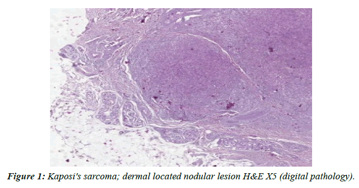
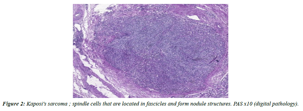
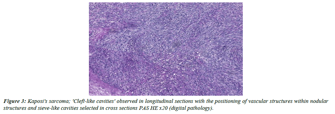
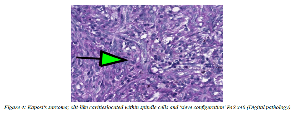
When we examined the slides sent for consultation and painted with H&E; we confirmed the diagnosis of 'Kaposi's sarcoma' put by our pathologist colleague (Figure 1-4).
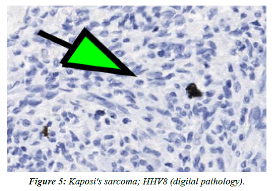
Why did we present this phenomenon? Goal; In order to be able to diagnose it in normal light microscopy, the 'kaposi sarcoma' factor (at least in most of them), which provides differentiation from lesions such as angiosarcoma, acroangiodermatitis, hobnail hemangioma, spindle cell hemangioma, Kaposi form hemangioendothelioma, which is included in the differential diagnosis of the case, is to draw attention to the difficulties and persistence in the emergence of the HHV8 immunohistochemical biomarker indicating the presence of CNHV. When our case reached us, we studied HHV8, ERG CD34, CD31, S-100, Melan A, Desmin, and SMA as immunohistochemical panels from the moment we determined the preliminary diagnosis of Kaposi sarcoma and the appropriate histopathological appearance. The HHV8 result, which we rely on the most in this panel, surprised us at first. Because we found a positivity that was very focal and barely discernible in individual cells (Figure 5).
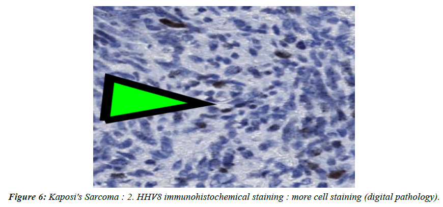
However; when we searched the literature, we observed that the same problem was encountered in the HHV8 (LANA) staining pattern, especially in the diagnosis of cutaneous kaposi sarcoma (Figure 6) [2].
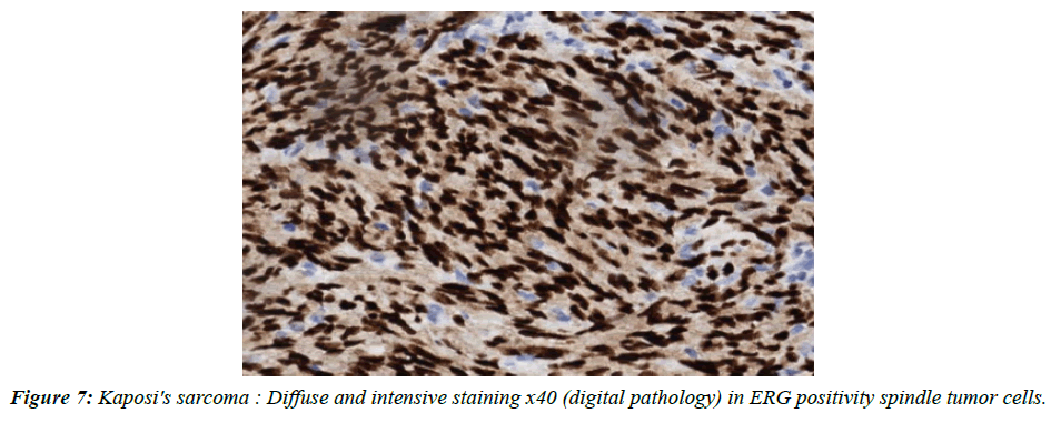
So we repeated our HHV8 IHK request. On subsequent HHV8 staining, more cells had positive staining. Ultimately; Together with ERG, CD31, CD 34 positivity, we presented our histopathological confirmed diagnosis to our clinician friends (Figure 7).
Conclusion & Discussion
Kaposi's sarcoma is a rare tumor in our country. However; developing technology, globalizing world, migrations have resulted in the positioning of people from different countries in our country in recent years. Therefore; we have started to see many diseases and tumours that we have rarely seen before much more often and we will continue to see them. In this context; we should design our diagnostic range accordingly and expand our differential diagnostic profile. Our case; it was a diagnosed consultation case. However; no immunohistochemical studies were conducted in the country where the diagnosis was made. So we insisted that it might be troublesome to confirm the diagnosis without seeing HHV8 positivity. Ultimately; our efforts yielded results and the treatment of our patient was started with the diagnosis we gave. By the way, the same patient; 8 more similar nodular lesions were detected and excised in the hand and forearm area and all of them were diagnosed as 'Kaposi's sarcoma' by us.
Kaposi's sarcoma; is a disease of endothelial cells of blood vessels and the lymphatic system. Despite its name, it is not classified as a sarcoma (a malignant tumor of mesanchymal origin) in many centres recently, as it is due to multicentre vascular hyperplasia. These centres are also located in; HHV8 in; suggests that it is necessary to produce the disease, but it is not enough [3].
The 4 general types of Kaposi's sarcoma have been described.
• Classic Kaposi's sarcoma: affects older men of Mediterranean and Central European descent and men in Sub-Saharan Africa. It is associated with diabetes mellitus, but not with HIV infection.
• Kaposi's sarcoma associated with the human immunodeficiency virus (HIV): it mainly affects men who have sex with men (MSM). Kaposi's sarcoma is one of the most common types of cancer in Uganda and Zambia, especially in children.
• Endemic or African Kaposi's sarcoma: occurs in children and young adults in some parts of Africa.
• Iatrogenic Kaposi's sarcoma: observed after drug therapy or transplantation that causes immune suppression.
In this group of 4; it has been shown that the form of kaposi sarcoma, which occurs especially when the immune system is suppressed, progresses at the mucosal level in a short time and leads to death with organ dysfunctions [4].
In another important study; it is stated that this opportunistic neoplasm, which is frequently observed in gay men, is closely related to the number of partners. At least that's what seems to be the case in San Francisco [5].
Incidence of Kaposi's sarcoma; In 1973 (before the AIDS epidemic) it was 1 in 100,000, in 1991 it increased to 33 per 100,000 at the peak of the AIDS epidemic, and then in 1998 HAART (after highly active antiretroviral treatment) fell to 2.8 per 100,000 [6].
Our aim in preparing this article is; not to repeat the known information as it is done in many places, but to introduce our 'Kaposi's sarcoma' phenomenon, which we encounter again years later due to a phenomenon, to our young friends, especially in the profession, and to draw attention to the fact that it is definitely added to the differential diagnosis and the difficulties in diagnosis, especially in some special cases.
Especially; Iatrogenic Kaposi's sarcoma; Kaposi's sarcoma is of particular concern for organ transplant patients in geographical areas associated with high levels of infection with herpes virus (CNH). In these places, most patients have the virus before transplantation; drugs cause the virus to reactivate. For the same reason, the use of corticosteroids and biologic substances such as rituximab infliximab and abatacept, which are prescribed for chronic inflammatory and autoimmune conditions, may also play an indirect role in the development of Kaposi sarcoma.
A final word; As our professors used to say in the medical school; Acting with the principle of 'there is no disease, there are patients', being open to innovations in today's world where the world is changing rapidly and the borders are getting closer, being persistent (not stubborn) during diagnosis or treatment seems to be the solution.
References
- Casper C, Carrell D, Miller KG, et al. HIV serodiscordant sex partners and the prevalence of human herpesvirus 8 infection among HIV negative men who have sex with men: baseline data from the EXPLORE Study. Sex Transm Infect. 2006; 82(3):229-35.
- Cassar O, Blondot ML, Mohanna S, et al. Human Herpesvirus 8 Genotype E in Patients with Kaposi Sarcoma, Peru. Emerg Infected Tooth. 2010; 16(9) :1459-62.
- Mesri EA, Cesarman E, Boshoff C. Kaposi’s sarcoma herpesvirus/Human herpesvirus-8 (KSHV/HHV8), and the oncogenesis of Kaposi’s sarcoma. Nat Rev Cancer. 2010;10(10):707.
- Paul Curtiss, Lauren C. Strazulla &Alwin E. Friedmen -Kien. Dermatol Treat. 2016; 6(465-470).
- McKusick L, Horstman W, Coates TJ. AIDS and sexual behavior reported by gay men in San Francisco. Am J Public Health. 1985;75(5):493-6.
- Eltom MA, Jemal A, Mbulaiteye SM, et al. Trends in Kaposi's sarcoma and non-Hodgkin's lymphoma incidence in the United States from 1973 through 1998. J Natl Cancer Inst. 2002;94(16):1204-10.
Indexed at, Google Scholar, Cross Ref
Indexed at, Google Scholar, Cross Ref
Indexed at, Google Scholar, Cross Ref
