Case Report - Journal of Bacteriology and Infectious Diseases (2022) Volume 6, Issue 2
Antimicrobials, disc diffusion, haramaya, salmonella, susceptibility as a treatment dystocia in cat.
Dese Kefyalew1 Abebe Fromsa2 Yemsrach Wubayehu2 Feyera Gemeda1*
1School of Veterinary Medicine, College of Agriculture and Veterinary Medicine, Jimma University, Ethiopia
2Department of Clinical Studies, College of Veterinary Medicine and Agriculture, Addis Ababa University, P. O. Box 34, Bishoftu, , Ethiopia
- Corresponding Author:
- Feyera Gemeda Dima
Department of Agriculture and Veterinary Medicine
Jimma University, Jimma, Ethiopia
E-mail: feyera.gemeda@ju.edu.et
Received: 03-Feb-2022, Manuscript No. AABID-22-53272; Editor assigned: 04-Feb-2022, PreQC No. AABID-21-53272(PQ); Reviewed: 03-Mar-2022, QC No. AABID-22-53272; Revised: 07-Mar-2022, Manuscript No. AABID-22-53272(R); Published: 07-Mar-2022, DOI:10.35841/aabid-6.2.107
Citation: Kefyalew D, Fromsa A, Wubayehu Y, et al. Antimicrobials, disc diffusion, haramaya, salmonella, susceptibility as a treatment dystocia in cat. J Bacteriol Infec Dis. 2022;6(2):107
Abstract
En bloc ovariohysterectomy is the technique that involves ovariohysterectomy before hysterectomy and removal of the neonates were performed as treatment of dystocia on a dog and cat. A 5-year-old local breed queen with a history of four parity presented Veterinary Teaching Hospital of Addis Ababa University College of Veterinary Medicine and Agriculture for having difficulty giving birth. As the owner complained that the queen gave birth to one dead kitten 14 hrs before then the queen straining frequently but with no progress. Before surgery, all physical parameters were evaluated. The queens’ rectal temperature was subnormal (37.80 c) attended with a distended abdomen, straining, and vaginal discharge of offensive odor. The surgery was performed aseptically under general anesthesia. A midline incision behind the umbilicus approximately 10 cm long midline incision was made to enable easy exteriorization of the uterus. The operative procedure for these cases including post-operative care and postoperative complication was described in a case report. The queen was recovered with minimal complication.
Keywords
Dystocia, En bloc ovariohysterectomy, Queen.
Introduction
Dystocia is the inability of the dam to expel the fetus at parturition through the birth canal without assistance. The incidence of dystocia in companion animals like bitch and queen are quite low but when it occurs it may constitute life-threatening situations. Dystocia occurs in approximately 5% of all parturitions in dogs and 3.3% to 5.8% of parturitions in queens [1]. Dystocia may be caused by maternal or fetal factors, and in some cases, a combination of both. Maternal factors include small pelvic size, abnormalities of the caudal reproductive tract, primary or secondary uterine inertia, malnutrition, parasitism, and other abnormalities of the uterus [2]. Fetal causes include fetal monsters, fetal oversize with the maternal pelvis, fetal malposition or mal-posture, and fetal death. The process of parturition in animals is divided into three stages. The first stage is characterized by in-apparent uterine contractions and progressive dilation of the cervix. The duration is usually 6–12 h in the bitch, and somewhat shorter in the queen. The queen may vocalize; show tachypnea and restlessness, and some will lie in the queening box and purr loudly. In some queens, the first stage of labor can pass without overt clinical signs. The second stage of labor includes the process of fetal expulsion through a fully dilated cervix. The queen usually has her first kitten within 1 h of the onset of stage two labor and subsequent kittens every 10–60 min, but this is quite variable. Parturition lengths of up to 10-12 hrs have been reported in the queen. The third stage of labor encompasses the time in which fetal membranes are expelled. Surgery may also be performed as an elective procedure in animals that have a history of dystocia or are predisposed to dystocia animals with pelvic fracture mal-union or a narrow pelvic canal. It has been suggested that hysterectomy should not be performed to avoid the additional stresses to the dam of and longer anesthetic time. En bloc ovariohysterectomy is an alternative to cesarean section in dogs and cats with dystocia [3].
Case History
The queen’s case history
A local breed queen of fourth parity was presented with a history of dystocia to the Veterinary Teaching Hospital of Addis Ababa University. The owner complained that the queen has already delivered one dead kitten 14 hours before it was presented to the hospital.
Physical examination
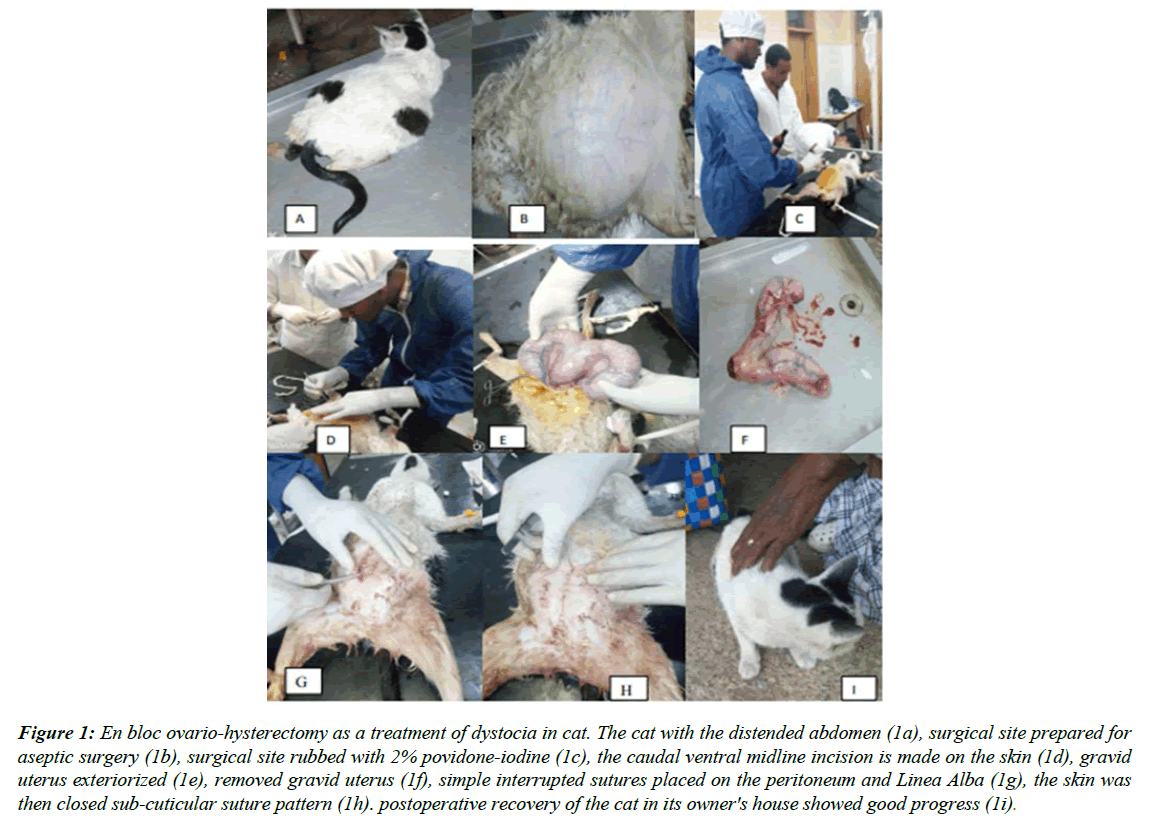
On general physical examination the animal was found frequently licking its perineal area. There was a vaginal discharge of offensive odor. The queen was frequently grunting and straining. A distended abdomen was also observed (Figure 1A). Upon clinical evaluation, the rectal body temperature was subnormal (37.80c) and the animal was dehydrated. On per vaginal examination, the birth canal was found moist with vaginal discharge that has a bad odor. Manual traction of the fetus that remained in the uterus was difficult due to the emphysematous fetus. Then a decision was made to perform emergency surgery by cesarean section was performed under general anesthesia to save the life of the queen.
Pre-operative animal preparation
The queen was sedated and put on a surgical table in dorsal recumbency and when the anesthetic took effect an IV catheter was put and connected to an IV fluid supply line containing lactated ringers solution administered at a surgical rate of 10 ml/kg/hr and delivered in calculated drop rate per second. The ventral midline abdominal area was shaved and washed with water and soap then scrubbed using savlon solution for aseptic surgery (Figure 1B), then it was finally rubbed with 2% povidone-iodine (Figure 1C). Anesthesia and Animal Control Atropine sulfate @ 0.3 mg/ kg was administered intramuscularly as premedical and general anesthesia was achieved by the combination of Xylazine Hydrochloride 0.3 mg/kg and Ketamine Hydrochloride 5mg/kg both drugs loaded into the same syringe and given intramuscularly to effect. When the pre-anesthetic took effect an IV catheter was put and connected to an IV fluid supply line containing lactated ringers solution administered at a surgical rate of 10 ml/kg/hr and delivered in a calculated drop rate per second. The patient was kept on the operation table and the surgery was done aseptically controlled under general anesthesia using a half dose of xylazine + ketamine combination. The queen was administered ceftriaxone at 20 mg /Kg intramuscularly before the operation as a prophylactic treatment to prevent potential infection of a surgical wound from the emphysematous fetus contained in the uterus.
Operative procedure
The surgical incision was made in the caudal midline approximately 2 cm behind the umbilicus. Approximately, 10-15cm long caudal ventral midline incision was made on the skin (Figure 1D). Bleeding from small cutaneous arteries was controlled by applying pressure using gauze and/or artery forceps. The subcutaneous tissues and fats were removed. Linea alba and peritoneum were incised layer by layer to exteriorize the uterus then the vascular pedicle containing the ovarian artery and vein was isolated by breaking down the mesovarium. No clamps were placed on the vessels. This procedure was repeated for the other ovary and the uterus was exteriorized (Figure 1E) and the ovarian pedicles were double ligated with 2-0 catgut suture. The broad ligament was broken down on both sides of the uterus to a point just cranial to the cervix. The cervix and vagina were palpated for fetuses, which were gently manipulated into the body of the uterus. The ovaries and uterus were removed by dividing between the clamps and were given to a team of assistants, who immediately opened the uterus, and seven dead emphysematous fetuses were appreciated. Then, the surgical site was inspected for evidence of bleeding (Figure 1F). The peritoneum and muscle layers were sutured with a simple interrupted pattern with catgut (1-0) (Figure 1G). The subcutaneous layer was sutured with a lockstitch pattern using catgut (1-0). The skin was then closed as a subcuticular suture pattern using catgut (1-0) (Figure 1H). The operation was successfully performed, the abdomen was closed routinely. Subcuticular sutures were preferred over skin sutures because of the proximity of the suture line to the mammary glands (Figure 1I).
Figure 1: En bloc ovario-hysterectomy as a treatment of dystocia in cat. The cat with the distended abdomen (1a), surgical site prepared for aseptic surgery (1b), surgical site rubbed with 2% povidone-iodine (1c), the caudal entral midline incision is made on the skin (1d), gravid uterus exteriorized (1e), removed gravid uterus (1f), simple interrupted sutures placed on the peritoneum and Linea Alba (1g), the skin was then closed sub-cuticular suture pattern (1h). postoperative recovery of the cat in its owner's house showed good progress (1i).
Discussion
En bloc ovariohysterectomy is performed particularly in animals for which cesarean section is indicated because of the risk of a difficult delivery [4]. In this case report, a similar technique of ovariohysterectomy was performed while the fetuses are still in the uterus. En bloc ovariohysterectomy may be performed whether the fetuses are alive or dead, and this method permanently blocks the reproductive ability without the need for a second operation [5]. The procedure was performed based on the interest of the owner. The survival rate of the newborn in dogs has been reported as 75% for cesarean sections performed by the en bloc technique and 92% for sections performed by the conventional method [6]. The queen was at fourth parity facing the difficulty of giving birth and the owner requesting for the queen be spayed for he needed no more kitten from his queen [7-9].
Conclusion
In this case report, all fetuses have died this way maybe the owner visited the assistant at the late stage of parturition. Reports have shown that ovariohysterectomy at the time of dystocia has stressed increased morbidity to the dam so, the use of en bloc ovariohysterectomy in cats and dogs had advantages of in minimal anesthetic time for the dam, and minimal potential for peritoneal contamination with uterine contents, which may occur during hysterotomy. En bloc ovariohysterectomy also provides an opportunity to affect population control in pets of clients who may not be able to afford to return for a second surgical procedure (ovariohysterectomy). En bloc ovariohysterectomy does not compromise the health of the dam.
Acknowledgements
The figures used in the case report belong to the author’s, captured in photograph while the author’s were conducting the surgical procedure. The case report was conducted at a certain Veterinary Teaching Hospital without any additional fund and prepared as surgical case report for learning and teaching of professionals across the world especially for veterinarians with your cooperation for processing and publication.
References
- Corbee R. Obesity in show cats. J Anim Physiol Anim Nutr (Berl) 98;2014:1075-80.
- Domoslawska A, Jurczak A, Janowski T. A one-foetus pregnancy monitored by Ultrasonography and progesterone blood levels in a German Shepherd bitch: a case report. Vet Med 56;2011:55-57.
- Farrow CS. Maternal-fetal evaluation in suspected canine dystocia: A radiographic prospective. Can Vet J 19;1978:24-26.
- Gunn-Moore D, Thrushfield M. Feline dystocia: prevalence and association with cranial comformation and breed. Vet Record 136(14);1995:350-53.
- Jackson P. Handbook of Veterinary Obstetrics. 2nd Edition. Saunders Elsevier and Philadelphia. 2004:2-8.
- Robbins MA, Mullen HS. En Bloc Ovariohysterectomy as a treatment for dystocia in dogs and cats. Vet Surg 23;1994:48-52.
- Probst CW, Webb AI. Cesarean section in the dog and cat: Anesthetic and surgical techniques, in Bojrab MJ (Edition): Current Techniques in Small Animal Surgery (2nd Edition). Phil- adelphia. PA, Lea & Febiger. 1983:346-51.
- Robbins M, Mullen H. En bloc ovariohysterectomy as a treatment for dystocia in dogs and cats. Vet Surg 23;2004:48-52.
- Sparkes A, Rogers K, Henley W, et al. A questionnairebased study of gestation, parturition and neonatal mortality in pedigree breeding cats in the UK. J Feline Med Surg, 8;2006:145-57.
Indexed at, Google Scholar, Cross Ref
Indexed at, Google Scholar, Cross Ref
Indexed at, Google Scholar, Cross Ref
Indexed at, Google Scholar, Cross Ref
