Imaging Brain - Biology & Medicine Case Reports (2017) Volume 1, Issue 1
A smooth brown snake-skin like hemispherical bulge in the stomach
- *Corresponding Author:
- Shiyang Ma, M.D.
Department of Gastroenterology The Second
Affiliated Hospital Xi’an Jiaotong University
Xi’an Shaanxi 710004 P.R. China
Tel: +86-29-87679335
Fax: +86-29-87679660
E-mail: shiyangma@163.com
Accepted date: July 26, 2017
Citation: Qiao L, Zhong W, Zhou M, et al. A smooth brown snake-skin like hemispherical bulge in the stomach. Biol Med Case Rep. 2017;1(1):7-8.
DOI: 10.35841/biology-medicine.1.1.7-8
Visit for more related articles at Biology & Medicine Case ReportsAbstract
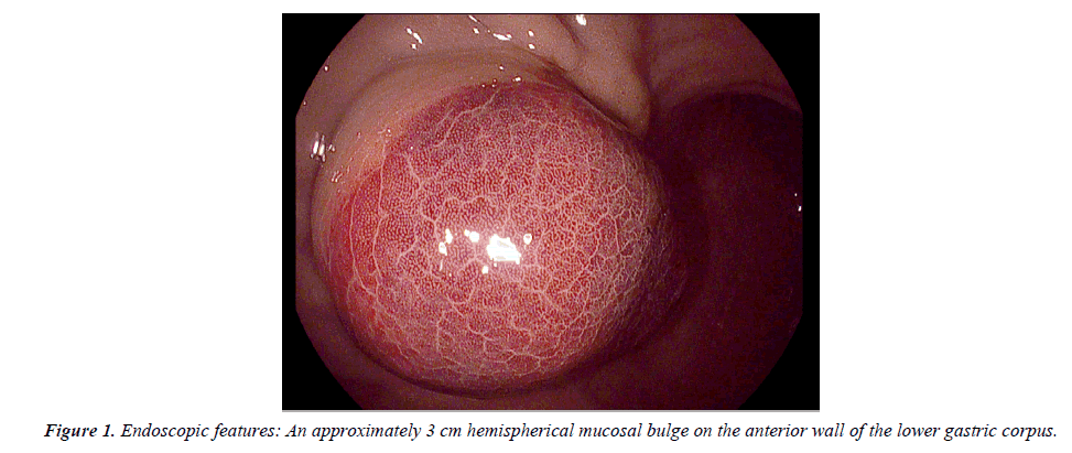
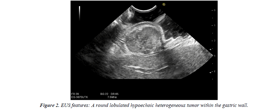
A 47-year-old Chinese female was admitted for epigastric discomfort and the gastric endoscopy showed an approximately 3 cm hemispherical mucosal bulge, with a smooth brown snake-skin like surface, on the anterior wall of the lower gastric corpus (Figure 1). The Endoscopic ultrasonography (EUS) showed he tumor originated from muscularis propria and presented as a round lobulated hypoechoic lesion with regular clear margins. There was an irregular echo-free necrosis in the lesion, with an unabundant blood supply and an integral serosa.
Keywords
Gastrointestinal stromal tumor, Endoscopic ultrasonography, Hyperemia of the mucosa.
Description
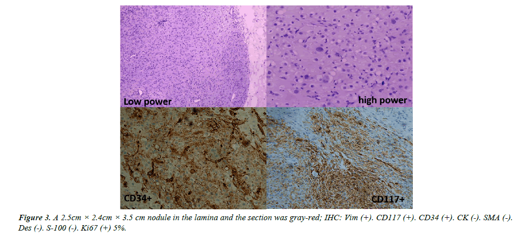
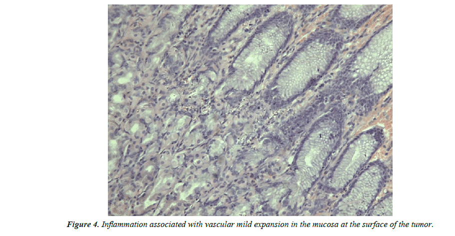
A 47-year-old Chinese female was admitted for epigastric discomfort and the gastric endoscopy showed an approximately 3 cm hemispherical mucosal bulge, with a smooth brown snake-skin like surface, on the anterior wall of the lower gastric corpus (Figure 1). The Endoscopic ultrasonography (EUS) showed the tumor originated from muscularis propria and presented as a round lobulated hypoechoic lesion with regular clear margins. There was an irregular echo-free necrosis in the lesion, with an unabundant blood supply and an integral serosa. The diameters of the largest cross-section were 23.5 × 16.6 mm (Figure 2). The dark red change may due to mild vascular expansion, the inflammatory cells infiltration or may also be associated with internal friction (Figure 3), tumor growth or increasing intracapsular pressure limiting the blood supply of mucosa, which could be in the early stage of an ulcer. Once the ulcer occurs or the internal pressure is released, the color of surrounding mucosa might be normal.
According the origination of the muscularis propria, and occupying the anterior wall with the maximum diameter of 23.5 mm, we prefered laporascopic resection. Finally the lesion was completely resected and proved a low-risk GIST without local or distant infiltration (Figure 4), thus the tyrosine kinase inhibitor imatinib is unnecessary.


