Case Report - Journal of Clinical Ophthalmology (2020) Volume 4, Issue 1
A case of Choroidal Hemangioma in pregnancy.
Krishna Nagaradh1*, Prarthana Gokarn21Sapthagiri Institute of Medical Sciences and Research Centre, Bengaluru, Karnataka, India
2Narayana Netralaya, Bengaluru, Karnataka, India
- Corresponding Author:
- Dr. Krishna Nagaradh
Sapthagiri Institute of Medical Sciences
and Research Centre Chikkasandra
Chikkabanavara Bengaluru Karnataka, India
E-mail: allwayskrish@gmail.com
Accepted date: January 27, 2020
Citation: Nagaradh K, Gokarn P. A case of Choroidal Hemangioma in pregnancy. J Clin Ophthalmol 2020;4(1):209-211.
Abstract
We report a 29 year old female gravid 1 with 28 weeks of gestation was referred by her obstetrician for blurred vision in her left eye from the past 3 days. On examination patient manifested a facial hemangioma over left face and lid. Fundus examination of left eye revealed a choroidal hemangioma which increased in size during the course of pregnancy. No intervention was done and the tumour regressed completely after pregnancy. Observation alone may be indicated in cases of choroidal hemangioma demonstrating sub-retinal fluid in pregnancy. Circumscribed choroidal hemangioma can manifest in Sturge-Weber syndrome rather typically found diffuse type.
Keywords
Choroidal hemangioma, Sturge weber syndrome, Pregnancy, Sub-retinal fluid.
Introduction
Choroidal hemangioma is an unusual, benign vascular hamartomas whose exact incidence is not clear. It usually presents between second to fourth decades of life. It can either be asymptomatic or present with varied symptoms like reduced vision, metamorphopsia, and photopsias [1]. Generally, they are orange-red elevated masses, which are found posterior to the equator. Lesions are usually solitary and unilateral [2]. Overlying subretinal fluid, serous retinal detachment and cystoid macular edema are common findings.
Choroidal hemangiomas usually exist as well demarcated circumscribed mass or as an ill-defined diffuse mass. Circumscribed tumors occur without any associated local or systemic anomalies. Diffuse choroidal hemangiomas are usually evident at birth, as a part of Sturge-Weber syndrome and are associated with ipsilateral cutaneous lesions, bilateral ocular signs and venous hemangioma of leptomeninges [3].
Sturge weber syndrome is a rare, sporadic neuro-cutaneous disorder which occurs in approximately one per 50,000 live births. The most significant, purely vascular anamoloy of the eye is a diffuse choroidal hemangioma, in contrast to wellcicumscribed choroidal hemangiomas seen in patients without the syndrome [4]. Only three cases of circumscribed choroidal hemangiomas have been reported in patients with SWS, both of which were bilateral in nature [5]. On our literature search there is very less data about the sturge weber syndrome associated with pregnancy.
Diagnosis of choroidal hemangioma is challenging as it can mimic serious intraocular lesions like choroidal melanoma, metastasis and other inflammatory lesions [6]. Our case study is the fourth case to be reported of a patient with SWS who developed a circumscribed choroidal hemangioma instead of the usual diffuse manifestation.
Case Report
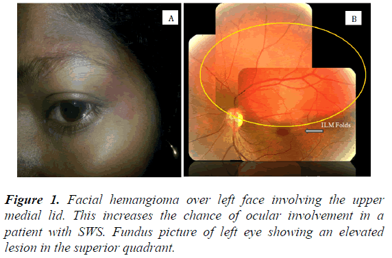
A 29 year old south Indian female gravid 1 with 28 weeks of gestation was referred by her obstetrician for blurred vision in her left eye from the past 3 days. No history of symptoms and signs of pre eclampsia and eclampsia. History of LASIK surgery in her right eye done 2 years back. On examination patient manifested a facial hemangioma over left face the upper medial lid. This increases the chance of ocular involvement in a patient with SWS. It consisted of dilated thin walled capillaries and venules, most of which are situated in the upper part of the reticular dermis. Her visual acuity at the time of presentation in right eye was 20/20 and in her left eye 20/60 N10 improving to 20/30. Intra ocular pressure was 16 mm of hg in both eyes.
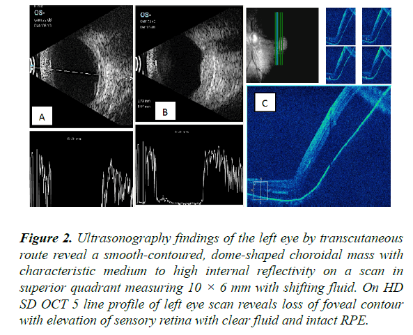
Findings from a dilated fundoscopic examination of left eye revealed normal vitreous. In the macula we found central pigmentary changes of the outer retinal layers as well as an area of elevation that encompassed the entire superior macula going beyond the arcade vessels, most likely corresponding to an exudative retinal detachment. Beneath this, she had a red elevated area with relatively sharp borders consistent with a choroidal hemangioma. She also had inferior ILM folds in the left eye (Figures 1 and 2).
Figure 2: Ultrasonography findings of the left eye by transcutaneous route reveal a smooth-contoured, dome-shaped choroidal mass with characteristic medium to high internal reflectivity on a scan in superior quadrant measuring 10 × 6 mm with shifting fluid. On HD SD OCT 5 line profile of left eye scan reveals loss of foveal contour with elevation of sensory retina with clear fluid and intact RPE.
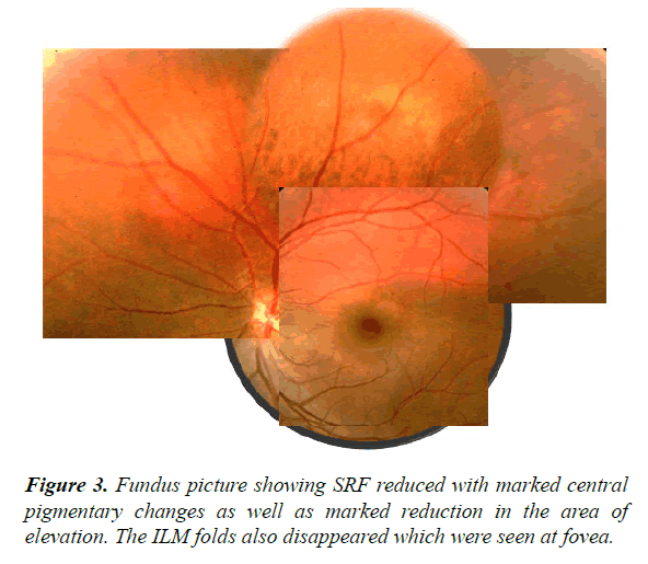
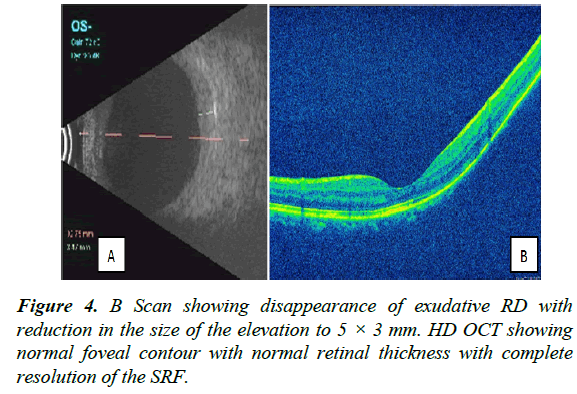
Patient was reviewed after 2 weeks, vision reduced from 20/60 to 20/120 and to 20/200 N36 on further follow-ups. Also there was an increase in the sub retinal fluid. No management was planned still at this stage and observation was the call. There after review was done every 2 weeks till delivery and until 3 months post-partum. Her vision in left eye had improved to uncorrected 20/30 N6 (Figures 3 and 4).
Discussion
Circumscribed choroidal hemangioma is considered rare. Jarrett et al diagnosed one circumscribed choroidal hemangioma for every 15 cases of choroidal melanoma [7]. In most clinical series on choroidal hemangioma, approximately 50% of tumors were circumscribed while 50% were diffuse tumors associated with Sturge-Weber syndrome.
In the Shields series, 67% of tumors were in the macula, 34% were between the macula and the equator, and no tumor was anterior to the equator. Mean tumor diameter was 6.7 mm, and mean tumor thickness was 3.1 mm. Serous retinal detachment and cystoid macular edema were reported in 81% and 17% of patients, respectively. All circumscribed choroidal hemangiomas reported by Witschel and Font, were located posterior to the equator, and subretinal fluid was noted in 47% of their patients [8]. Our case had tumour located in the posterior pole just above the fovea extending superiorly for size 11 × 9 mm with ILM folds at the fovea.
Little is known regarding the behavior of choroidal hemangioma in pregnancy. There is one report of leakage from a circumscribed choroidal hemangioma in pregnancy, and the fluid resolved following full term normal delivery [9]. In another report of three patients with choroidal hemangioma, exudative retinal detachments developed during the third trimester of pregnancy [10], two cases showed complete resolution after delivery. The third patient suffered from total retinal detachment and secondary glaucoma, and finally required enucleation [10].
Regarding safety of FFA in pregnancy the correct answer no one knows. Most retinal experts don ’t perform fluorescein angiography on pregnant women. The risk of congenital malformations or spontaneous abortions is probably low with fluorescein angiography but no one can say that it’s completely safe. Also our literature review showed case reports with completed resolution of fluid after delivery. So we decided to remain safe and observe without any ancillary tests or any treatment till her delivery and intention was to treat the patient with photodynamic therapy (PDT) after delivery. Our patient showed complete resolution of subretinal fluid with vision recovery after delivery.
We attribute this resolution, to normalcy of altered hemodynamic state during pregnancy owing to hormonal changes during pregnancy which may result in engorgement of vascular networks within the hemangioma, leading to increased transudation of fluid into the sub-retinal space.
Conclusion
Observation alone may be indicated in cases of hemangioma demonstrating sub-retinal fluid in pregnancy with keeping in mind that spontaneous resolution always is not a rule. Circumscribed choroidal hemangioma can manifest in Sturge- Weber syndrome rather than typically found diffuse type.
Financial Interest
None financial disclosures.
Conflicts of Interest
Conflicts of interest none with any of the authors.
References
- Turell ME, Singh AD. Vascular tumours of retina and choroid: Diagnosis and treatment. Middle East Afr J Ophthalmol. 2010;17:191-200.
- Boixadera A, García-Arumí J, Martínez-Castillo V, et al. Prospective clinical trial evaluating the efficacy of photodynamic therapy for symptomatic circumscribed choroidal hemangioma.Ophthalmology. 2009;116:100-105.
- Zablocki G, Hagedorn C, Brady KD. Choroidal Hemangioma: Central scotoma in the right eye. EyeRounds.org. 2007.
- Cheung D, Grey R, Rennie I. Circumscribed choroidal haemangioma in a patient with Sturge Weber syndrome. Eye (Lond). 2000;14:238-40.
- Sturge WA. A case of partial epilepsy, apparently due to a lesion of one of the vasomotor centres of the brain. Transactions of the Clinical Society of London. 1879;12:162.
- Shields JA, Stephens RF, Eagle RC Jr, et al. Progressive enlargement of a circumscribed choroidal hemangioma. A clinicopathologic correlation. Arch Ophthalmol. 1992;110:1276-8.
- Jarrett WH 2nd, Hagler WS, Larose JH, et al. Clinical experience with presumed hemangioma of the choroid: Radioactive phosphorus uptake studies as an aid in differential diagnosis. Trans Sect Ophthalmol Am Acad Ophthalmol Otolaryngol. 1976;81:862-70.
- Witschel H, Font RL. Hemangioma of the choroid. A clinicopathologic study of 71 cases and a review of the literature. Surv Ophthalmol. 1976;20:415-31.
- Pitta C, Bergen R, Littwin S. Spontaneous regression of a choroidal hemangioma following pregnancy. Ann Ophthalmol. 1979;11:772-4.
- Cohen VM, Rundle PA, Rennie IG. Choroidal hemangiomas with exudative retinal detachments during pregnancy. Arch Ophthalmol. 2002;120:862-4.


