Research Article - Biomedical Research (2017) Volume 28, Issue 7
The analysis of the effect of intervertebral disc on the lateral bending of the spine in deformed thorax model
Jin-duo Ye1, Chun-yue Zhang1, Wei-hong Zhong1, Bo-lun Liu1, Ji-fu Liu2 and Chun-qiu Zhang1*1Tianjin Key Laboratory of the Design and Intelligent Control of the Advanced Mechatronical System, School of Mechanical Engineering, Tianjin University of Technology, Tianjin, 300384, PR China
2Department of Thoracic Surgery, General Hospital of Beijing Command, Beijing, PR China
- *Corresponding Author:
- Chun-qiu Zhang
Tianjin Key Laboratory of the Design and Intelligent Control of the Advanced Mechatronical System
School of Mechanical Engineering, Tianjin University of Technology, PR China
Accepted date: December 28, 2016
Abstract
Objective: To assess the functions of intervertebral discs on the lateral bending of the spine in deformed thorax model, and to improve the reliability of the minimally invasive correction surgery for treating pectus excavatum.
Background: Clinical outcomes demonstrate that improvement of scoliosis after surgery correction does not meet expected results if the influences of intervertebral discs have not been taken into account during pre-surgery numerical simulation of pectus excavatum with scoliosis.
Method: Two thorax models were reconstructed based on the computed tomographic (CT) data, which included the deformed (pectus excavatum with scoliosis) models with and without intervertebral discs. In order to study the effects of these discs on the lateral bending of the spine, a 40 mm correcting displacement was applied on the sagittal plane of the sternum of the deformed thorax models.
Results: The result of the lateral displacement of the spine was smaller in the deformed thorax model with discs (2.1 mm) than without discs (2.7 mm). After correction, the Cobb angle of the deformed thorax with discs was 13°, whereas the Cobb angle was 11° in the model without discs. Actually the clinical correction result was 15°.
Conclusion: Intervertebral discs decrease the lateral bending of the spine in the deformed thorax model. The computed results demonstrate that intervertebral discs play a critical role in the lateral bending of the spine of deformed thorax models. Thus, the influence of intervertebral discs should be considered in numerical model and simulation of minimally invasive correction surgery of pectus excavatum with scoliosis.
Keywords
Pectus excavatum with scoliosis, Thoracic intervertebral disc, Numerical model, Numerical simulation
Introduction
The NUSS procedure has been widely used in children as a minimally invasive procedure for treating pectus excavatum [1-4]. During this correction procedure, a deformed sternum and ribs may contribute to significant pull on the spine, which may results in an uncertain surgery result and/or aggravation of scoliosis, which have to lead an early termination of the operation [5-10].
There is problem need to be further improved in numerical analysis, though recently, establishing finite-element models have been widely used to profile the deformation in pectus excavatum with scoliosis. The numerical simulation of the based model can be used to predict orthopedic results to get the best treatment results [11-14]. In 2010, Nagasao et al. prior to surgery, established thorax models with beam elements while simulating correction procedures for 25 patients with asymmetric pectus excavatum [15]. The effects of NUSS surgery on the spine were determined by comparing the thorax images pre- and post-NUSS surgery. The results suggested that the predicted spine shape of the numerical simulation was consistent with the surgical result in several cases, whereas in some cases the spine exacerbated deformity after the surgery while the simulation results were positive [15]. Therefore, it suggest that the discrepancy between simulation results and clinical results may lie in the underestimation of the effects of thoracic discs, in fact, the simulation model above exaggerated the spine bending stiffness. Moreover, there are few researches that have assessed the effects of thoracic discs on the lateral bending of the spine.
Discs, acting as the connective tissue between two vertebrae, somehow determine the biomechanical properties of the spine. Thus, we hypothesized that intervertebral discs influence the lateral bending of the spine in deformed thorax model. In order to elucidate the underlying biomechanical effects of the discs on the lateral bending of the spine, we tested this hypothesis by simulating correction procedures on a deformed thorax of pectus excavatum with scoliosis. Our study provides a foundation for further study and design of correction surgery plans for pectus excavatum, and it is of significance for improving the reliability of the minimally invasive correction surgery of pectus excavatum with scoliosis.
Materials and Methods
Model construction
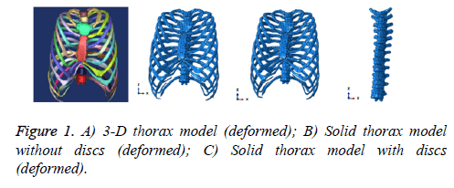
Computed tomography (CT) images of thorax of a patient with pectus excavatum with scoliosis were provided by General Hospital of Beijing Command. In the patient with pectus excavatum with scoliosis, the sternum inward concave to the right side with a depth of 40 mm, and the spine bowed to the left with a Cobb angle of 20°. The 3-D solid model of the deformed thorax was reconstructed in Mimics (Mimics 10.0; Materialise Technologies, Leuven, Belgium) (Figure 1A). The models were imported into Geomagic (Geomagic studio 10.0; GeomagicInc, North Carolina, USA) for point cloud processing, and finally imported into ABAQUS(ABAQUS 6.11) to establish the assembled solid model (Figure 1B). Model 1-b was the thorax model without discs. Accordingly, we propose a feasible way to construct the thorax model with discs. In ABAQUS, columns that were thicker than the discs were produced in the gaps between vertebrae, and the disc between two vertebrae was obtained by the Boolean calculations of subtraction based on the upper and lower vertebra as well as the column. Thorax models constructed by using this approach are shown in Figure 1C.
Numerical simulation
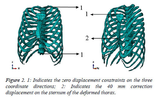
The two models were pre-processed in ABAQUS, respectively. The sternums, ribs, and thoracic vertebrae were connected by coupling nodal displacements (same displacement for adjacent nodes), while the discs and the vertebrae were connected by Boolean calculation of glue. Subsequent meshes were added to the models. We build models using proper material parameters of the model [12,13,15] and boundary conditions based on surgery method. Table 1 shows the settings of the element parameters. The boundary conditions were defined as follows: the upper surface of the first thoracic vertebra, the lower surface of the 12th thoracic vertebra as well as the interfaces between the sternums and the collar bones were defined as the zero displacement constraints on the three coordinate directions (Figure 2). Additionally, because rib displacement is large during the correction procedure, we took into account during each calculation the potential for geometric nonlinear. For the simulation on the deformed thorax, a -40 mm correction displacement (along the Y direction) was applied to the lower part of the second sternum. We compared the lateral bending of the spine, in terms of both displacement field and stress distribution in the deformed thorax models with or without discs.
| Name | Elastic modulus (MPa) | Poisson ratio |
|---|---|---|
| Sternum | 450 | 0.42 |
| Thoracic vertebra | 450 | 0.42 |
| Rib | 162 | 0.45 |
| Intervertebral disc | 10 | 0.48 |
Table 1: Parameters of the model material.
Results
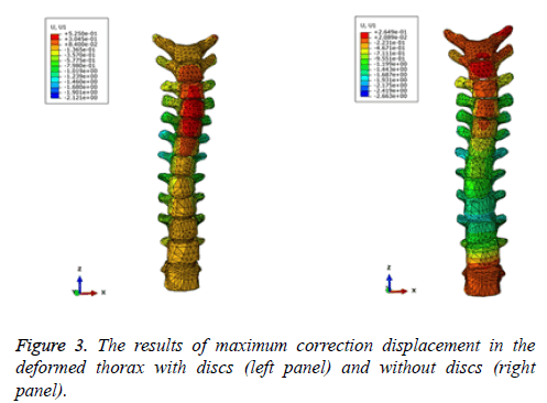
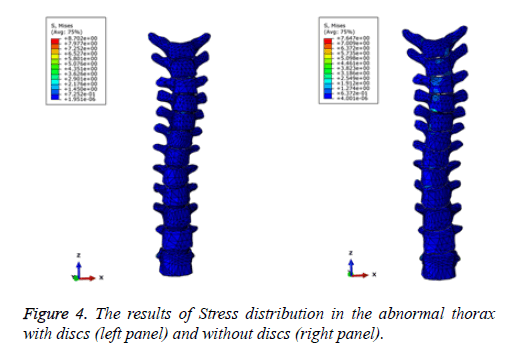
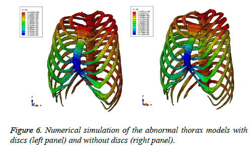
The results of the maximum correction displacement (X) of the spine were shown in Figure 3. Which show the spine deformation occurred on the lateral bending in the thorax, and the maximum spine displacement values (-X) in the models with and without discs were 2.1 mm (at 5th to 7th vertebrae) and 2.7 mm (at 5th to 9th vertebrae). The results showed that discs have influence on lateral displacement of the spine. The results of stress distribution on the spine are presented in Figure 4. We can find the maximum correction stress on the spine of the deformed thorax models with and without discs were 8.7 MPa (at the 6th vertebra) and 7.6 MPa (at the 4th vertebra). From computing results, the discs have effect on stress distribution in the spine.
Validation of simulation results
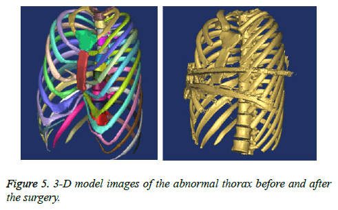
The simulation results of the deformed thorax models with and without discs were compared with the correction result after surgery. Surgeons implanted two correcting bars at the lower part of the second sternum (the concaved position of pectus excavatum) to correct pectus excavatum. Figure 5 shows 3-D solid models of the deformed thorax before and after surgery, while Figure 6 presents the simulation result of correction in the thorax models with and without discs. The comparison of the clinical correction result in these three models is presented in Table 2.
| Name | Post-surgery | Numerical simulation result | |
|---|---|---|---|
| (With discs) /Relative error |
(Without discs) /Relative error |
||
| Deformation of the sternum (Y/mm) | -41.03 | -41.43/0.975% | -41.32/0.707% |
| Cobb angle of the spine | 15° | 13°/13.333% | 11°/26.667% |
Table 2: Post-surgical deformity in comparison with the simulation results.
Before surgery, the patient had pectus excavatum that was inward concave to the right side with a depth of 40 mm, while the spine bowed to the left with a Cobb angle of 20°. As shown in Table 2, the simulation results from the thorax models with and without discs showed the deformation of the sternum were both close to the clinical result, which also demonstrated small relative errors. Cobb angles improved 13° and 11° in simulation models with and without discs, respectively, while the respective relative errors were 13.3% and 26.7% as compared with clinical results.
Overall, simulation results of improved scoliosis based on the model with discs was similar to surgical results, which was accompanied by smaller relative error compared to simulation results based on the model without discs. The research results suggested that the discs have an important influence on the lateral bending of spine in deformed thorax model. The relative error was calculated with the following formula:

where A is the post-surgical value and B is the simulated value.
Discussion
In pre-surgery numerical simulation, the new function of the intervertebral discs has been confirmed that in the correction of pectus excavatum with scoliosis, intervertebral discs absorb the lateral bending of the spine, which do not meet with the fact that intervertebral discs may increase the lateral bending of the spine. The results of the paper demonstrate that there is small difference in lateral bending of the spine between the numerical results based on models with intervertebral discs and the results of post-surgery correction, which is in contrast to simulation results based on models without intervertebral discs. The paper suggest that numerical simulation of minimally invasive correction of pectus excavatum with scoliosis using the numerical models with intervertebral discs is more consistent with clinical results than the numerical models without discs. Moreover, although the result which considered the intervertebral discs was closed to the clinical results, but there are still some errors after consideration the discs. It suggests that there may be other factors affecting the lateral bending of the spine of deformed thorax model, in which there may be the intercostal muscles. There are several subtypes of pectus excavatum with scoliosis. The position and the depth of pectus excavatum as well as the direction and the height of scoliosis all exert direct influences on the deformation of the spine. Therefore, this mechanism of this influence need to be elucidated in future studies in this field.
Conclusion
1. Two numerical models of deformed thorax with or without intervertebral discs have been constructed in the paper, while studying the deformation patterns of the lateral bending of the spine under loading conditions by numerical simulation. The simulation results show that intervertebral discs decrease the lateral bending of the spine of deformed thorax. These findings potentially explain the reason that the orthopedic surgery for pectus excavatum with scoliosis fails to meet with the clinical results when surgical plans has been designed based on simulation models without considering the influence of intervertebral discs.
2. In comparing our prediction results with clinical results, we demonstrate that the prediction results based on the simulation using the thorax model of pectus excavatum with scoliosis was consistent with the real-world correction result when the prediction model takes into account intervertebral discs. However, the simulation using the thorax model of pectus excavatum with scoliosis without considering intervertebral discs shows large error compared to surgical results.
3. The results of the paper suggest that the effects of intervertebral discs should be taken into account in the numerical model when simulating the correction of pectus excavatum with scoliosis in order to predict clinical results more accurately.
Availability of data and supporting materials
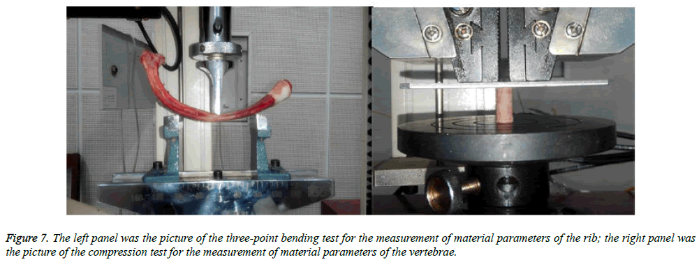
Confirmation method of the material parameters of human rib and vertebrae in the paper, it took three-point bending test for the test specimens of animal (pig) ribs and compression test for vertebral test specimens to obtain the Elastic modulus (E) and Poisson's ratio (μ), as it is shown in Figure 7. The testing machine was adopted WDW-10 computer-controlled electronic universal testing machine, made by Changchun Kexin Test Instrument Co. Ltd. The material parameters of rib was E=148 MPa and μ=0.45. The material parameters of vertebra was E=318M Pa and μ=0.43. With reference of animal experiments and related literatures, this paper determined the material parameters of ribof human thorax model was E=162 MPa and μ=0.45, the material parameters of vertebra was E=450 MPa and μ=0.42. Referring to the vertebrae material parameters, the sternum material parameters wasset as E=450 MPa and μ=0.42. The setting of the material parameters of intervertebral disc were referred to related literatures [16,17], E=10 MPa and μ= 0.48.
Acknowledgement
The project was supported by the National Natural Science Foundation of China (No.11372221) and partly supported by the National Natural Science Foundation of China (11432016 , 11402171,11672208).
References
- Krasopoulos G, Dusmet M, Ladas G. Nuss procedure improves the quality of life in young male adults with pectusexcavatum deformity. Euro J Cardio Thoracic Surgery 2006; 29: 1-5.
- Frick SL. Scoliosis in children with anterior chest wall deformities. Chest SurgerClin North Am 2000; 10: 427-436.
- Felts E, Jouve JL, Blondel B. Child pectusexcavatum: correction by minimally invasive surgery. Ortho Traumatol Surgery Res 2009; 95: 190-195.
- Protopapas AD, Athanasiou T. Peri-operative data on the Nuss procedure in children with pectusexcavatum: independent survey of the first 20 years' data. J Cardiothoracic Surgery 2008; 3: 1.
- Nuss D, Kelly RE, Croitoru DP. A 10-year review of a minimally invasive technique for the correction of pectusexcavatum. J Pediatric Surgery 1998; 33: 545-552.
- Jaroszewski DE, Fonkalsrud EW. Repair of pectus chest deformities in 320 adult patients: 21 year experience. Annal Thoracic Surgery 2007; 84: 429-433.
- Engum S, Rescorla F, West K. Is the grass greener? Early results of the NussPprocedure. J Pediatric Surgery 2000; 35: 246-251.
- Pilegaard HK, Licht PB. Routine use of minimally invasive surgery for pectusexcavatum in adults. Annal Thoracic Surgery 2008; 86: 952-956.
- Johnson WR, Fedor D, Singhal S. Systematic review of surgical treatment techniques for adult and pediatric patients with pectusexcavatum. J Cardiothoracic Surgery 2014; 9: 1.
- Nagasao T, Miyamoto J, Tamaki T. Stress distribution on the thorax after the Nuss procedure for pectusexcavatum results in different patterns between adult and child patients. J Thoracic Cardiovascular Surgery 2007; 134: 1502-1507.
- Zhong WH, Ye JD, Liu JF. Numerical Simulation and Clinical Verification of the Minimally Invasive Repair of PectusExcavatum. Open Biomed Eng J 2014; 8: 147.
- Ye J, Wang Z, Zhang C. The numerical modeling to the orthopedic of thorax with pectusexcavatum. 2011 International Conference on Communications and Networks (CECNet) IEEE, 2011.
- Wei Y, Sun D, Liu P. Pectusexcavatumnussorthopedic finite element simulation. 2010 3rd International Conference on Biomedical Engineering and Informatics. IEEE 2010.
- Wei Y, Shi Y, Wang H, Gao Y. Simulation of Nussorthopedic for pectusexcavatum. 2nd International Conference on Biomedical Engineering and Informatics. IEEE, 2009.
- Nagasao T, Noguchi M, Miyamoto J. Dynamic effects of the Nuss procedure on the spine in asymmetric pectusexcavatum. J Thoracic CcardiovascularSurgery 2010; 140: 1294-1299.
- Schmidt H, Heuer F, Drumm J. Application of a calibration method provides more realistic results for a finite element model of a lumbar spinal segment. ClinBiomech 2007; 22: 377-384.
- Schmidt H, Heuer F, Wilke HJ. Interaction between finite helical axes and facet joint forces under combined loading. Spine 2008; 33: 2741-2748.
