Research Article - Journal of Clinical Dentistry and Oral Health (2022) Volume 6, Issue 6
RETROSPECTIVE EVALUATION OF SEPARATED INSTRUMENTS TO THE TYPE OF USED FILE , TOOTH TYPE, CANAL TYPE AND PORTION OF CANAL- DATA ANALYSIS IN UNIVERSITY SETTING
Kavalipurapu Venkata Teja* and Sharon Keziah
Department of Conservative dentistry and endodontics, Saveetha Dental College and Hospitals, Saveetha Institute of Medical and Technical Sciences, Saveetha University, Chennai, India
- Corresponding Author:
- Teja KV
Department of Conservative dentistry and endodontics
Saveetha Dental College and Hospitals
Saveetha Institute of Medical and Technical Sciences
Saveetha University, Chennai
E-mail: venkatatejak.sdc@saveetha.com
Received: 28-October-2022, Manuscript No. AACDOH-22-80355; Editor assigned: 31-October-2022, PreQC No. AACDOH-22-80355 (PQ); Reviewed: 15-November-2022, QC No. AACDOH-22-80355; Revised: 18-November-2022, Manuscript No.AACDOH-22-80355 (R); Published: 25-November-2022, DOI: 10.35841/aacdoh-6.6.128
Citation: Sharon Keziah V, Teja KV. Retrospective evaluation of separated instruments to the type of used file, tooth type, canal type and portion of canal- data analysis in university setting. J Clin Dent Oral Health. 2022;6(6):128
Abstract
Instrument separation is a common occurrence during the endodontic treatment due to the use of hand or rotary files, Gates Glidden burs, lentulo, pluggers, spreaders made of different materials like NiTi, stainless steel. The mishap of instrument separation is a frustrating situation for the endodontist as it prevents access to the apex and impedes full length instrumentation and obturation of the root canal. Aim: This study evaluated the separated instruments to the type of used file, tooth type, canal type. Materials and method: There were a total of 78 instrument separation cases from June 2020- March 2021 that were evaluated. Patient details like age, gender, tooth type, canal type, portion of canal, type of instrument and method of retrieval were included in the study. The collected Data was analysed using SPSS. Results: The odds for rotary instrument separation were higher than the hand instruments with 57%. The probability of separating a file in the coronal third was more compared to the apical and middle third. The highest percentage of instrument separation in the mandibular posteriors with 36%. Furthermore, the odds of file separating in a single canal (38%) and mesiobuccal canal (19%) were higher. There was no significant difference (p>0.05) in instrument separation between type of instrument and portion of canal involved.
Keywords
Hand instrument, Instrument separation, Root canal, Dental innovation.
Introduction
Endodontic treatment has been made much easier by the revolutionization of rotary files with super flexibility [1]. The use of these rotary files has significantly improved the cleaning and shaping process of the canals and reduced the chair time of the operator significantly [2]. However these instruments do have their disadvantages. The biggest hindrance for the cleaning and shaping process in root canal treatment is the separation of the endodontic instruments [3]. This has a potential impact on the outcome of the treatment. Instrument separation during endodontic treatment is a common accident involving rotary or hand files with rotary files having a higher incidence.
There are many factors leading to the instrument separation such as anatomic variations, frequency of usage, speed and torque of rotation [4]. Regarding the anatomical variation of the canals, previous literature has shown if the canal anatomy is complicated, there are more chances of instrument separation. It has been reported that the frequency of instrument separation is more in molars. The mesial canals of mandibular molars are more commonly affected [4,5]. Also the apical third is more prone to instrument separateion than coronal and middle third. Certain studies assessed the curvature of the root canal, where the separation frequency is directly proportional to curvature with 58% in intensely curved canals [6]. The cyclic flexural fatigue resistance decreases with increased use. Regarding the speed, there have been previous reports that during the high speed of rotation there is an increase in the prevalence of instrument separation compared to low speed of rotation [7]. A higher rate of instrument separation was observed when speed of 300–350 rpm was used. However other studies have stated that there is no effect of speed on file separation. Based on the torque of rotation, if high torque is used there are higher chances of instrument separation due to the deformation of the file [8]. Files in smaller diameter and files in acutely curved canals have more tendency to separate in the root canal. .Severe curvature is one of the main causes for instrument breakage because of cyclic fatigue [9].
The approach of cases with a separated instrument can be conservative or surgical. The conservative approach involves a) Bypass of the separated instrument, b) Removal of the fragment, c) Instrumentation and obturation coronally to the fragment. Regarding the removal of a separated instrument, a number of techniques have been developed [10]. Ultrasonics, along with the operative microscope, is the most effective and reliable method for removing a separated endodontic instrument from a root canal [11]. The possibility of successful removal depends on: the level of separation whether coronal, middle or apical third, location in relation curvature, the type of instrument, length, the degree of canal curvature and the tooth type [3]. There are several complications that may occur during the management of a separated instrument. Separation of the ultrasonic tip or file used for bypassing , further separation of the fragment, perforation, extrusion of the file into periapical tissues and excessive temperature rise in periodontal tissues are some of the complications during the retrieval [12]. The mishap of instrument separation created a frustrating situation for the dentist as it prevented access to the apex and most of the time impedes full length instrumentation and obturation of the root canal [12-17]. As a result of this mishap, the level of difficulty is increased. Of the certain case augments, while the tooth healing is challenged [18-23]. The aim of this study is to evaluate the separated instruments to the type and taper of used file, tooth type, canal type and radius of the curvature.
Material and methods
This study was designed as a retrospective cross sectional observational study analysing all the cases of instrument separation. The data of patients between the age of 18 years and above who underwent instrument retrieval from June 2020 to March 2021 for instrument separation were included in the study. The records with incomplete medical documentation, replication of results in different time periods with improper clinical photographs or diagnosis were excluded from the study. Patient details like age, gender, type of instrument, tooth type, canal type, canal portion and method of retrieval were studied. The collected Data was described as frequency distribution and percentile. Statistical analysis was performed using Statistical Package for the Social Sciences, version 22(SPSS). Descriptive analysis were based on quantitative variables and frequencies for categorical variables. A Chi square test was applied to determine the significance between groups. p value< 0.05 was considered to be statistically significant with a confidence interval of 95%.
Results
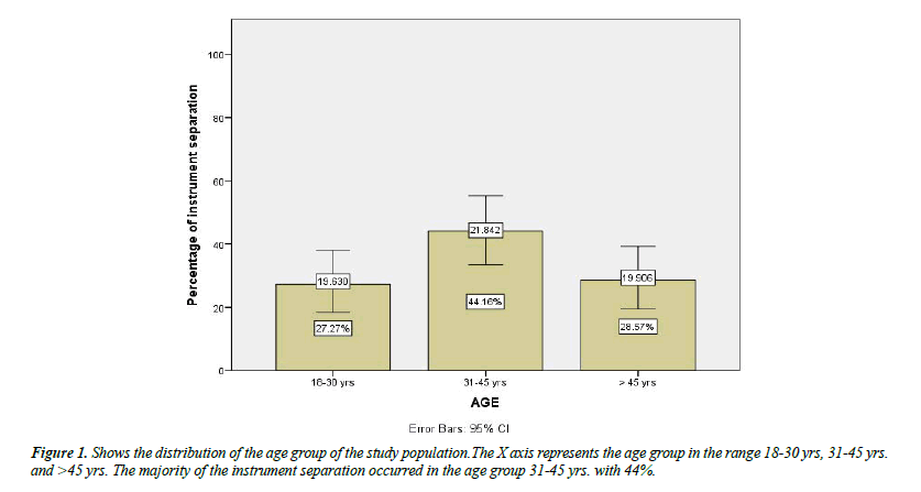
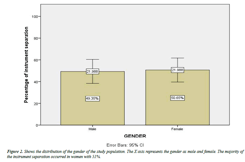
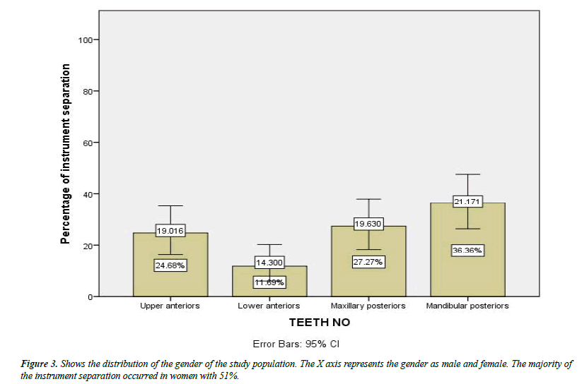
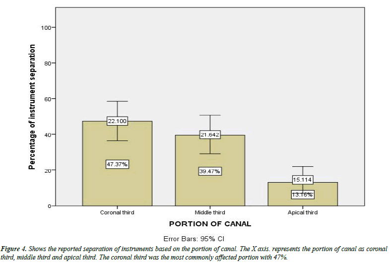
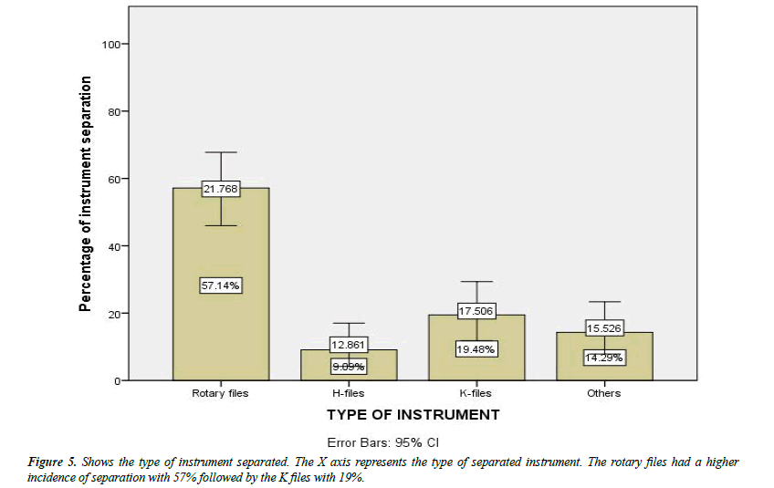
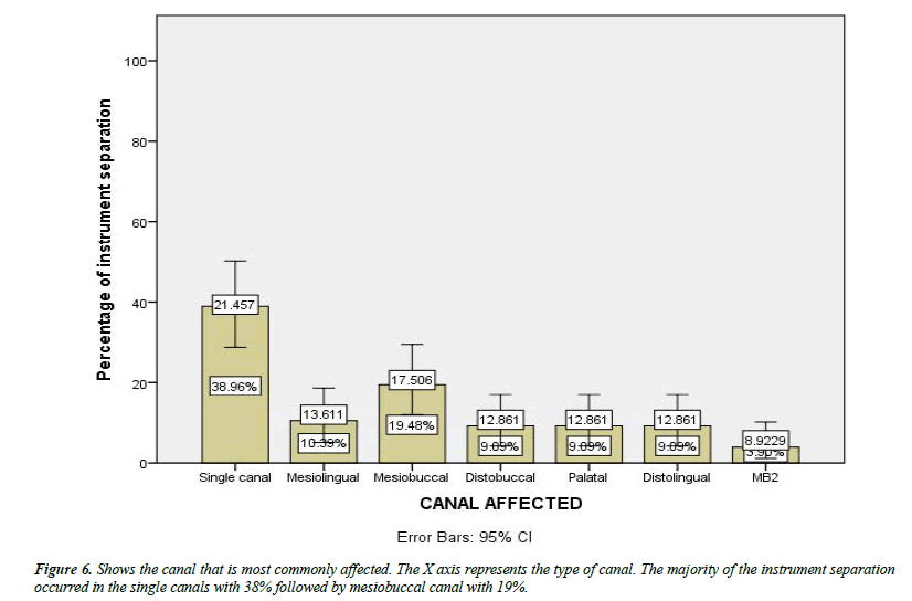
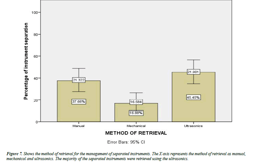
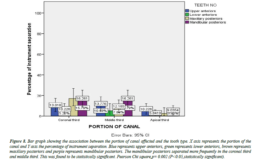
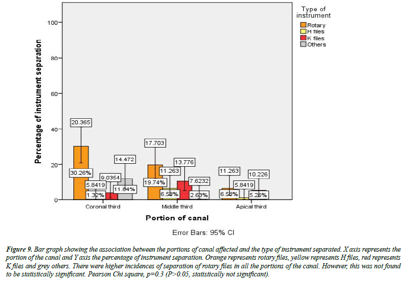
There were a total of 78 separated instruments observed in this study. The majority of them were present in the age group 31-45 yes (Figure 1). There were 49% males included in the study and 51% were females (Figure 2). When hand and rotary instruments were combined, the mandibular posterior teeth accounted for 36% of the separated files, followed by maxillary posterior teeth with 19% (Figure 3). The likelihood of instrument separation in the current study was higher in the coronal portion with 47% than in the middle and apical third (Figure 4). The instrument separation incidence of rotary files and K files was 57% and 20%, respectively (Figure 5). Of the 78 separated instruments, 38% separated in a single canal followed by 19% in the mesiobuccal canal (Figure 6). According to the method of retrieval of separated instruments, the majority (45%) were retrieved with the help of ultrasonics (Figure 7). The association between the tooth type and the portion of the canal in which the instrument separated was found to be statistically significant, with p=0.002 (Figure 8). The association between the type of instrument and portion of the canal in which the instrument separated showed no significant association, p =0.3 (Figure 9)
Figure 8:Bar graph showing the association between the portion of canal affected and the tooth type. X axis represents the portion of the canal and Y axis the percentage of instrument separation. Blue represents upper anteriors, green represents lower anteriors, brown represents maxillary posteriors and purple represents mandibular posteriors. The mandibular posteriors separated more frequently in the coronal third and middle third. This was found to be statistically significant. Pearson Chi square,p= 0.002 (P<0.05,statistically significant).
Figure 9:Bar graph showing the association between the portions of canal affected and the type of instrument separated. X axis represents the portion of the canal and Y axis the percentage of instrument separation. Orange represents rotary files, yellow represents H files, red represents K files and grey others. There were higher incidences of separation of rotary files in all the portions of the canal. However, this was not found to be statistically significant. Pearson Chi square, p=0.3 (P>0.05, statistically not significant).
Discussion
Cleaning and shaping is an important part of endodontic therapy. The errors during the procedure such as zipping, transportation, and perforations can be significantly reduced with NiTi rotary instruments than the stainless steel ones. However, NiTi rotary instruments do have their own disadvantages like all the other instruments and that is intracanal breakage or instrument separation. The results of this present study have observed that the odds of a separated rotary instrument are greater when compared to hand instruments. Previous literature has reported an overall incidence of NiTi rotary file separation as 1.67 %. The mandibular posterior teeth have a higher incidence of instrument separation. Similar results were seen in previous studies where the incidence of 2.5% in mandibular molars are considered. This could be attributed to the complex root canal anatomy of the molars.
The primary cause of NiTi separation has been because of torsional and fatigue failure of NiTi. During torsional failure, the tip of the instrument is stuck or locked in the root canal while the remaining instrument further continues to rotate therefore causing the instrument separation. Flexural failure occurs because of cyclic fatigue at the center of the greatest curvature of the root canal. The file separates at the fracture line without unwinding of flutes. The cause of instrument separations in the current study might be because of flexural failures. This assumption is supported by the fact that in multirooted teeth the instrument separations occurred more in the mesiobuccal canals which are known for their greater curvatures. It has previously been reported that rotary NiTi instruments usually fracture at the midpoint of their curvature within simulated root canals, so it was surprising to find that most of the instruments in our study separated in the coronal third of canals.
However this could be attributed to the small sample siz. Contradictory to these findings, a previous study observed that the apical third was most commonly involved in the canal, where canals typically curve and have their smallest diameters. The probability of instrument separation in the mesiobuccal canal of molars was almost three times greater when compared to the distobuccal canal. The MB canal of the mandibular molar is known for its greater curvature and therefore a higher incidence, similar to the present study. Additionally, the mesial canals of mandibular molars coalesce to form a major foramen in about 49% of the cases. These type of canal should be instrumented only to the point where it coalesce the main canal because instrumenting till its full working length will force the file to navigate the abrupt curvature probably leading to instrument separation.
It should be understood that no definite conclusions could be given regarding incidence of instrument separation. There are various factors that affect the separation rate. These include: the times the instrument has been used, the techniques used, manual glide path, the size to which the canals are enlarged, torque, the combination of instruments used and the depth of knowledge regarding configuration of the root canals.The present study limits the comprehensive data assessment on the specific topic. Future studies could assess more on the level of instrument separation to the success of the therapy rendered [14-31].
Conclusion
The results of the current study show that rotary instruments have a greater tendency to separate in root canals than hand instruments. The instruments are more likely to separate in the single canals and mesiobuccal canals of multirooted teeth and also in the coronal third of the root canals. Prognosis of the tooth is not affected by presence of the separated instrument but by the presence of an infection of the root canal at the time of separation. Hence more care should be taken during the endodontics therapy to minimise such incidences.
Acknowledgements
This research was done under the supervision of the Department of Endodontics, Saveetha dental College and Hospital. We sincerely show gratitude to the corresponding guide who provided insight and expertise that greatly assisted the research.
Conflict of Interest
There was no potential conflict of interest.
Source of Funding
Saveetha Dental College & Hospitals, Saveetha Institute of Medical and Technical Sciences, Saveetha University Kumar Automobiles, Chennai, India.
References
- Suter B, Lussi A, Sequeira P. Probability of removing fractured instruments from root canals. Int Endod J. 2005;38(2):112-23.
- https://dspace.cuni.cz/handle/20.500.11956/171651
- Lambrianidis T. Management of fractured endodontic instruments: A clinical guide. Springer. 2017; 28.
- Baumann MA, Roth A. Effect of experience on quality of canal preparation with rotary nickel-titanium files. Oral Surg Oral Med Oral Pathol Oral Radiol Endod. 1999;88(6):714-718.
- Al-Fouzan KS. Incidence of rotary profile instrument fracture and the potential for bypassing in vivo. Int Endod J. 2003;36(12):864-867.
- Hülsmann M, Herbst U, Schäfers F. Comparative study of root-canal preparation using lightspeed and quantec SC rotary NiTi instruments. Int Endod J. 2003;36(11):748-756.
- Ankrum MT, Hartwell GR, Truitt JE. K3 Endo, ProTaper, and profile systems: Breakage and distortion in severely curved roots of molars. J Endod. 2004 ;30(4):234-237.
- Fishelberg G, Pawluk JW. Nickel-titanium rotary-file canal preparation and intracanal file separation. Compend Contin Educ Dent. 2004; 25(1):17.
- Camilleri J. Endodontic materials in clinical practice. John Wiley and Sons. 2021;320.
- Knowles KI, Hammond NB, Biggs SG, et al. Incidence of instrument separation using light speed rotary instruments. J Endod. 2006; 32(1):14-16.
- Orstavik D, editor. Essential endodontology: Prevention and treatment of apical periodontitis. John Wiley and Sons. 2020.
- Wolcott S, Wolcott J, Ishley D, et al. Separation incidence of protaper rotary instruments: A large cohort clinical evaluation. J Endod. 2006;32(12):1139-1141.
- Yang Y, Ye H, Zhao C, et al. Value added immunoregulatory polysaccharides of hericium erinaceus and their effect on the gut microbiota. Carbohydr Polym. 2021;262:117668.
- PradeepKumar AR, Shemesh H, Nivedhitha MS, et al. Diagnosis of vertical root fractures by cone-beam computed tomography in root-filled teeth with confirmation by direct visualization: A systematic review and meta-analysis. J Endod. 2021;47(8):1198-1214.
- Teja KV, Ramesh S. Is a filled lateral canal–A sign of superiority?. J Dent Sci. 2020;15(4):562.
- Narendran K, MS N, Sarvanan A. Synthesis, characterization, free radical scavenging and cytotoxic activities of phenylvilangin, a substituted dimer of Embelin. Indian J Pharm Sci. 2020;82(5):909-912.
- Gupta R, Kaur N, Sharma V, et al. Prevalence and risk factors associated with traumatic dental injuries among 12–15 year old school going children, Mathura city. J Indian Assoc Public Health Dent. 2021;19(1):76.
- Sawant K, Pawar AM, Banga KS, et al. Dentinal microcracks after root canal instrumentation using instruments manufactured with different NITI alloys and the saf system: A systematic review. Appl Sci. 2021;11(11):4984.
- Bhavikatti SK, Karobari MI, Zainuddin SL, et al. Investigating the antioxidant and cytocompatibility of Mimusops elengi Linn extract over human gingival fibroblast cells. IJERPH. 2021;18(13):7162.
- Maiese K. Sirtuin biology in medicine: Targeting new avenues of care in development, aging, and disease. Academic Press 2021.
- Ezhilarasan D. MicroRNA interplay between hepatic stellate cell quiescence and activation. Eur J Pharmacol. 2020;885:173507.
- Romera A, Peredpaya S, Shparyk Y, et al. Bevacizumab biosimilar BEVZ92 versus reference bevacizumab in combination with FOLFOX or FOLFIRI as first-line treatment for metastatic colorectal cancer: A multicentre, open-label, randomised controlled trial. Lancet Gastroenterol Hepatol. 2018;3(12):845-855.
- Raj R K. ß-Sitosterol-assisted silver nanoparticles activates Nrf2 and triggers mitochondrial apoptosis via oxidative stress in human hepatocellular cancer cell line. J Biomed Mater. 2020;108(9):1899-1908.
- Li B, Moriarty TF, Webster T, et al. Racing for the surface: Antimicrobial and interface tissue engineering. Morgantown, WV, USA. Springer; 2020.
- Lichtfouse E, Schwarzbauer J, Robert D, et al. Environmental chemistry for a sustainable world: Remediation of air and water pollution. Springer Science and Business Media. 2011.
- Karobari MI, Basheer SN, Sayed FR, et al. An in vitro stereomicroscopic evaluation of bioactivity between Neo MTA Plus, Pro Root MTA, BIODENTINE & glass ionomer cement using dye penetration method. Materials. 2021;14(12):3159.
- Rohit Singh T, Ezhilarasan D. Ethanolic extract of Lagerstroemia speciosa (L.) Pers., induces apoptosis and cell cycle arrest in HepG2 cells. Nutr Cancer. 2020;72(1):146-56.
- Shukla AK. Nanoparticles and their biomedical applications. Springer. 2021;286.
- Vijayashree Priyadharsini J. In silico validation of the non-antibiotic drugs acetaminophen and ibuprofen as antibacterial agents against red complex pathogens. J Periodontol. 2019;90(12):1441-1448.
- Uma Maheswari TN, Nivedhitha MS, Ramani P. Expression profile of salivary micro RNA-21 and 31 in oral potentially malignant disorders. Braz Oral Res. 2020;34.
- Chaturvedula BB, Muthukrishnan A, Bhuvaraghan A, et al. Dens invaginatus: A review and orthodontic implications. Br Dent J. 2021;230(6):345-50.
Indexed at, Google Scholar, Cross Ref
Indexed at, Google Scholar, Cross Ref
Indexed at, Google Scholar, Cross Ref
Indexed at, Google Scholar, Cross Ref
Indexed at, Google Scholar, Cross Ref
Indexed at, Google Scholar, Cross Ref
Indexed at, Google Scholar, Cross Ref
Indexed at, Google Scholar, Cross Ref
Indexed at, Google Scholar, Cross Ref
Indexed at, Google Scholar, Cross Ref
Indexed at, Google Scholar, Cross Ref
Indexed at, Google Scholar, Cross Ref
Indexed at, Google Scholar, Cross Ref
Indexed at, Google Scholar, Cross Ref
Indexed at, Google Scholar, Cross Ref
