Research Article - Journal of Clinical Dentistry and Oral Health (2022) Volume 6, Issue 3
Most common shade used in direct veneer.
Ashwin Shravan Kumar, Vigneshwar T*Department of Conservative Dentistry and Endodontics, Saveetha Dental College, Saveetha Institute of Medical and Technical Sciences (SIMATS), Saveetha University, Chennai, India
- *Corresponding Author:
- Vigneshwar T
Department of Conservative Dentistry and Endodontics
Saveetha Dental College
Saveetha Institute of Medical and Technical Sciences (SIMATS)
Saveetha University, Chennai, India
E-mail: vigneshwart.sdc@saveetha.com
Received: 21-Apr-2022, Manuscript No. AACDOH-22-61372; Editor assigned: 23-Apr-2022, PreQC No. AACDOH-22-61372 (PQ); Reviewed: 7-May-2022, QC No. AACDOH-22-61372; Revised: 10-May-2022, Manuscript No. AACDOH-22-61372 (R); Published: 17-May-2022, DOI:10.35841/aacdoh- 6.3.114
Citation: Kumar AS, Vigneshwar T. Most common shade used in direct veneer. J Clin Dentistry Oral Health 2022; 6(3):114
Abstract
Introduction: Veneers with direct resins are one of the common treatment options for clinical applications following the developments in adhesive and restorative dentistry in recent years. These restorations are applied on prepared tooth surfaces or even without any preparation, with an adhesive agent and a composite resin material directly in a single visit in the dental clinic. If done properly, the aesthetic outcomes of direct composite veneers are very satisfactory in addition to superior optical and physical properties. In recent history these restorations were thought to be temporary alternatives to indirect ceramic veneers; however, they are no longer named 'day savior fillings' today. These restorations are called minimally invasive, functional and long-lasting 'direct aesthetic restorations' that perfectly emulate natural dental tissues even in anterior area. Discolorations of teeth or restorations, dental malformations or mal-positions, diastemas, crown fractures and abrasive or erosive defects are some examples of up-to-date indications of direct composite veneers. In this study we would evaluate the common shares used in direct veneer restoration. Materials and Methods: The clinical records of all direct veneering cases during the period between 1 January 2020, and 1 January 2021 were. Shade selection was taken into account. Gender and age of the patients were also included in the study. Results: It can be seen that the majority of the direct veneers were placed in relation to maxillary central incisors and the most commonly used shade was found to be A2. How much color change can be achieved under such thin restorations depends on the thickness, color and opacity level of the veneer and the luting resin. Understanding the parameters that guide the modulation of hue, chroma and value will allow the clinician to modify a set color by mixing different shades in varying proportions until the desired color is finally achieved. Conclusion: Within the limitations of the study it can be said that most common shade used among both the gender was found to be A2 for direct veneer restoration
Keywords
Ecofriendly, Novel technique, Innovative technique, Guide, Shade, Gender.
Introduction
Veneers with direct resins are one of the common treatment options for clinical applications following the developments in adhesive and restorative dentistry in recent years. These restorations are applied on prepared tooth surfaces or even without any preparation, with an adhesive agent and a composite resin material directly in a single visit in the dental clinic [1]. If done properly, the aesthetic outcomes of direct composite veneers are very satisfactory in addition to superior optical and physical properties [2]. In recent history these restorations were thought to be temporary alternatives to indirect ceramic veneers; however, they are no longer named 'day savior fillings' today. These restorations are called minimally invasive, functional and long-lasting 'direct aesthetic restorations' that perfectly emulate natural dental tissues even in anterior area [3-6]. Discolorations of teeth or restorations, dental malformations or mal-positions, diastemas, crown fractures and abrasive or erosive defects are some examples of up-to-date indications of direct composite veneers.
Enamel hypoplasia is a developmental malformation generally resulting in poor aesthetics, tooth sensitivity, malocclusion and predisposition to dental caries. Direct composite veneer restorations where the whole labial surface is covered with resin, are good treatment options in such cases [7]. In recent years direct composite veneers have been compared to indirect ceramic veneers and declared as weak in relation to resistance to fractures and discolorations. However, the fact that everyone misses is whether these direct resin restorations were done correctly or not? Direct composite veneers have indications and contraindications like almost all other dental procedures which can be listed as proper occlusion, lateral and protrusive movements, correct shade analysis, effective isolation, good adhesion, effective polishing, [8-10]. Our team has extensive knowledge and research experience that has translated into high quality publications [11-30]. The aim of this article is to evaluate the common shares used in direct veneer restoration.
Materials and Methods
The study was done as a retrospective, single centered study. Ethical approval was obtained from the Institutional Ethical Committee (Ethical approval number. SDC/ SIHEC/ 2020/ DIASDATA/ 0619-0320). We reviewed case records of the data of patients who had direct veneers in anterior teeth. Incomplete data were excluded. Age, gender and the site of veneer were collected. These data were cross verified with photographs and radiographs. The collected data were analysed using SPSS statistical software. Descriptive statistics (percentage, mean, SD) and inferential test (Chi-square test) were done appropriately.
Results and Discussion
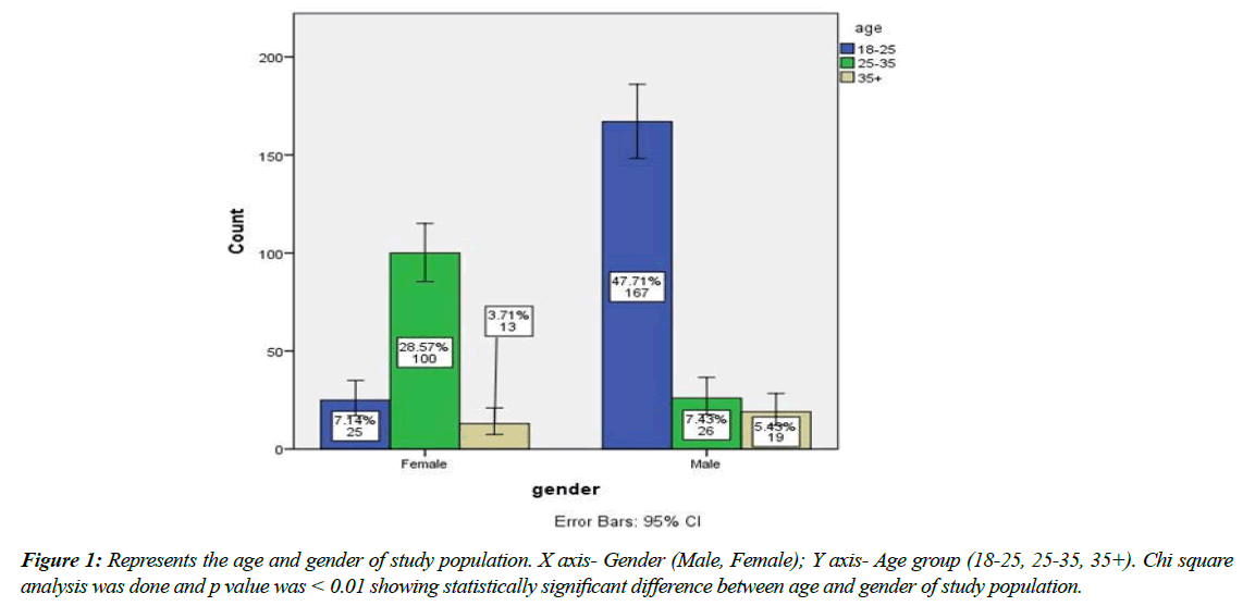
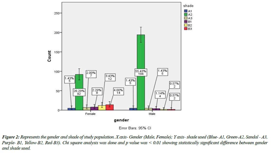
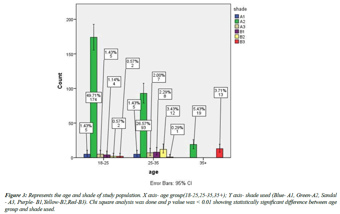
From Figure 1 it can be seen that the majority of the participants were males and were of age group 18-25. Also from Figure 2 and Figure 3 it can be said that the most common shade used was A2. Mazzota et al in his study stated that the majority of shade matching processes done resulted with A2 enamel shade and B2 dentin shade as most preferred. Our study has been in accordance with that. Also Budiprammanna et al., stated that A2 shade has been widely preferred in anterior aesthetic restorations of any forms. It can be seen that majority of the direct veneers were placed in relation to maxillary central incisors and the most commonly used shade was found to be A2. The direct–indirect composite veneer technique was introduced in the 1990’s as a means to heat-temper composites in partial and full veneers [31-33].
Figure 2: Represents the gender and shade of study population. X axis- Gender (Male, Female); Y axis- shade used (Blue- A1, Green-A2, Sandal - A3, Purple- B1, Yellow-B2, Red-B3). Chi square analysis was done and p value was < 0.01 showing statistically significant difference between gender and shade used.
Figure 3: Represents the age and shade of study population. X axis- age group(18-25,25-35,35+); Y axis- shade used (Blue- A1, Green-A2, Sandal - A3, Purple- B1,Yellow-B2,Red-B3). Chi square analysis was done and p value was < 0.01 showing statistically significant difference between age group and shade used.
In the direct–indirect technique, using similar shade selection and layering techniques that are used for the direct technique, the clinician applies a light-cured composite material to the tooth, with or without tooth preparation, without any adhesion. The composite is then shaped to a primary anatomic form with slight excess, and then light-cured. After that, the partially polymerized restoration is carefully removed (lifted) from the non-retentive, non-bonded tooth surface, heat-tempered extraorally chairside, and finished and polished to final macro and micro anatomy. After shade try-in and confirmation of the overall fit and esthetics, the veneer is bonded to the preparation using a resin-based luting agent.
One of the greatest benefits of the direct–indirect technique is that minor color changes can be realized through the use of luting resins of varying shades and degrees of opacity. This is very beneficial especially when trying to match a single veneer to adjacent, untreated teeth, and when doing multiple units and the patient's input is needed to determine the color matching alternative that best pleases her or him. How much color change can be achieved under such thin restorations depends on the thickness, color and opacity level of the veneer and the luting resin. Understanding the parameters that guide the modulation of hue, chroma and value will allow the clinician to modify a set color by mixing different shades in varying proportions until the desired color is finally achieved [34].
Conclusion
Within the limitations of the study it can be said that most common shade used among both the gender was found to be A2 for direct veneer restoration.
Funding
This research is supported/contributed/funded by Saveetha Institute of Medical And Technical Sciences, Saveetha Dental College and Hospitals, Saveetha University sponsored by Kumaran TV centre and Furniture pvt.ltd.
Conflict of Interest
Nil
Acknowledgements
Nil
References
- Barrantes JCR, Araujo E Jr, Baratieri LN. Clinical Evaluation of Direct Composite Resin Restorations in Fractured Anterior Teeth. Odovtos - Int J Dental Sci. 2015:47.
- Shikder AZ, Shomi K, Saki N, et al. Clinical evaluation of direct composite resin and indirect micro ceramic composite resin restorations in class-I cavity of permanent posterior teeth. Int J Hum Health Sci. 2019;3(02):109-5.
- Alvanforoush N, Palamara J, Wong RH, et al. Comparison between published clinical success of direct resin composite restorations in vital posterior teeth in 1995–2005 and 2006–2016 periods. Aus Dental J. 2017;62(2):132-45.
- Mykyievych NI. Efficiency Of Treatment Of Hard Tissues Defects Of Lateral Teeth With Direct And Indirect Restorations Made Of Composite Materials: Comparative Clinical Evaluation. Ukrainian Dental Almanac. 2018;(1):40-6.
- Cetin AR, Unlu N. One-year clinical evaluation of direct nanofilled and indirect composite restorations in posterior teeth. Dental Mat J. 2009;28(5):620-6.
- Blum IR. Composite restorations in anterior teeth: fundamentals and possibilities. Br Dental J. 2006;201(5):313.
- Strazzi-Sahyon HB, Passos Rocha E, Gonçalves Assunção W, et al. Influence of Light-Curing Intensity on Color Stability and Microhardness of Composite Resins. Int J Periodon Restor Dent. 2020;40(1).
- Winkler MM, Katona TR, Paydar NH. Finite element stress analysis of three filling techniques for class V light-cured composite restorations. J Dental Res. 1996;75(7):1477-83.
- Muthukrishnan L. Imminent antimicrobial bioink deploying cellulose, alginate, EPS and synthetic polymers for 3D bioprinting of tissue constructs. Carbo Poly. 2021;260:117774.
- PradeepKumar AR, Shemesh H, Nivedhitha MS, et al. Diagnosis of vertical root fractures by cone-beam computed tomography in root-filled teeth with confirmation by direct visualization: a systematic review and meta-analysis. J Endo. 2021;47(8):1198-214.
- Chakraborty T, Jamal RF, Battineni G, et al. A review of prolonged post-COVID-19 symptoms and their implications on dental management. Int J Environ Res Public Health. 2021;18(10):5131.
- Muthukrishnan L. Nanotechnology for cleaner leather production: a review. Environ Chem Lett. 2021;19(3):2527-49.
- Teja KV, Ramesh S. Is a filled lateral canal–A sign of superiority?. J Dent Sci. 2020;15(4):562.
- Narendran K, MS N, Sarvanan A. Synthesis, Characterization, Free Radical Scavenging and Cytotoxic Activities of Phenylvilangin, a Substituted Dimer of Embelin. Ind J Pharmac Sci. 2020;82(5):909-12.
- Reddy P, Krithikadatta J, Srinivasan V, et al. Dental caries profile and associated risk factors among adolescent school children in an urban South-Indian city. Oral Health Prev Dent. 2020;18(1):379-86.
- Sawant K, Pawar AM, Banga KS, et al. Dentinal Microcracks after Root Canal Instrumentation Using Instruments Manufactured with Different NiTi Alloys and the SAF System: A Systematic Review. App Sci. 2021;11(11):4984.
- Bhavikatti SK, Karobari MI, Zainuddin SL, et al. Investigating the Antioxidant and Cytocompatibility of Mimusops elengi Linn Extract over Human Gingival Fibroblast Cells. Int J Enviro Res Public Hea. 2021;18(13):7162.
- Karobari MI, Basheer SN, Sayed FR, et al. An In Vitro Stereomicroscopic Evaluation of Bioactivity between Neo MTA Plus, Pro Root MTA, BIODENTINE & Glass Ionomer Cement Using Dye Penetration Method. Mat. 2021;14(12):3159.
- Rohit Singh T, Ezhilarasan D. Ethanolic extract of Lagerstroemia Speciosa (L.) Pers., induces apoptosis and cell cycle arrest in HepG2 cells. Nutr Cancer. 2020;72(1):146-56.
- Ezhilarasan D. MicroRNA interplay between hepatic stellate cell quiescence and activation. Euro J Pharmacol. 2020;885:173507.
- Romera A, Peredpaya S, Shparyk Y, et al. Bevacizumab biosimilar BEVZ92 versus reference bevacizumab in combination with FOLFOX or FOLFIRI as first-line treatment for metastatic colorectal cancer: a multicentre, open-label, randomised controlled trial. Lancet Gastroenterol Hepatol. 2018;3(12):845-55.
- Raj R K. β‐Sitosterol‐assisted silver nanoparticles activates Nrf2 and triggers mitochondrial apoptosis via oxidative stress in human hepatocellular cancer cell line. J Biomed Mat Res Part A. 2020;108(9):1899-908.
- Vijayashree Priyadharsini J. In silico validation of the non‐antibiotic drugs acetaminophen and ibuprofen as antibacterial agents against red complex pathogens. J Periodontol. 2019;90(12):1441-8.
- Priyadharsini JV, Girija AS, Paramasivam A. In silico analysis of virulence genes in an emerging dental pathogen A. baumannii and related species. Archiv Oral Biol. 2018;94:93-8.
- Uma Maheswari TN, Nivedhitha MS, Ramani P. Expression profile of salivary micro RNA-21 and 31 in oral potentially malignant disorders. Braz Oral Res. 2020;34.
- Gudipaneni RK, Alam MK, Patil SR, et al. Measurement of the maximum occlusal bite force and its relation to the caries spectrum of first permanent molars in early permanent dentition. J Clini Pediatr Dent. 2020;44(6):423-8.
- Chaturvedula BB, Muthukrishnan A, Bhuvaraghan A, et al. Dens invaginatus: a review and orthodontic implications. Br Dent J. 2021;230(6):345-50.
- Kanniah P, Radhamani J, Chelliah P, et al. Green synthesis of multifaceted silver nanoparticles using the flower extract of Aerva lanata and evaluation of its biological and environmental applications. Chem Select. 2020;5(7):2322-31.
- Behnaz M, Dalaie K, Mirmohammadsadeghi H, et al. Shear bond strength and adhesive remnant index of orthodontic brackets bonded to enamel using adhesive systems mixed with tio 2 nanoparticles. Dental Press J Orthodon. 2018 ;23:43-e1.
- Kensche A, Dähne F, Wagenschwanz C, et al. Shear bond strength of different types of adhesive systems to dentin and enamel of deciduous teeth in vitro. Clin oral Inves. 2016;20(4):831-40.
- Triwardhani A, Budipramana M, Sjamsudin J. Effect of different white-spot lesion treatment on orthodontic shear strength and enamel morphology: In vitro study. J of Int Oral Health. 2019;12(2):120-8.
- Kulkarni G, Mishra VK. Enamel Wetness Effects on Microshear Bond Strength of Different Bonding Agents (Adhesive Systems): An in vitro Comparative Evaluation Study. J Conte Dental Prac. 2016;17(5):399-407.
- Savian TG, Oling J, Soares FZ, et al. Vital Bleaching Influences the Bond Strength of Adhesive Systems to Enamel and Dentin: A Systematic Review and Meta-Analysis of In Vitro Studies. Opera Dent. 2021;46(2):E80-97.
- Paradella TC, Fava M. Bond strength of adhesive systems to human tooth enamel. Braz Oral Res. 2007;21(1):4-9.
Indexed at, Google Scholar, Cross Ref
Indexed at, Google Scholar, Cross Ref
Indexed at, Google Scholar, Cross Ref
Indexed at, Google Scholar, Cross Ref
Indexed at, Google Scholar, Cross Ref
Indexed at, Google Scholar, Cross Ref
Indexed at, Google Scholar, Cross Ref
Indexed at, Google Scholar, Cross Ref
Indexed at, Google Scholar, Cross Ref
Indexed at, Google Scholar, Cross Ref
Indexed at, Google Scholar, Cross Ref
Indexed at, Google Scholar, Cross Ref
Indexed at, Google Scholar, Cross Ref
Indexed at, Google Scholar, Cross Ref
Indexed at, Google Scholar, Cross Ref
Indexed at,Google Scholar, Cross Ref
Indexed at, Google Scholar, Cross Ref
Indexed at, Google Scholar, Cross Ref
Indexed at, Google Scholar, Cross Ref
Indexed at, Google Scholar, Cross Ref


