Case Report - Otolaryngology Online Journal (2016) Volume 6, Issue 2
Carcinoma Glottis with Isolated Pelvic Bone Metastasis
- *Corresponding Author:
- Mohsin Khan
J N Medical College Hospital, ALIGARH, India
E-mail: khan.mohsin_1786@yahoo.com
Received: March 05, 2016; Accepted: March 15, 2016; Published: March 20, 2016
Abstract
Introduction: Bony metastasis from primary glottic carcinoma is a rare occurrence. Case report: 62 year old male presented with history of hoarseness of voice, swelling in left side of neck and swelling in right lower back. Physical examination revealed enlarged level ii and level IV left cervical lymph nodes. Direct laryngoscopy and biopsy established it a case of moderately differentiated squamous cell carcinoma glottis. X ray pelvic bone revealed osteolytic destruction right superior iliac crest, bone scan showed increased tracer uptake in right iliac bone and cytology from this bony lesion showed metastatic squamous cell carcinoma with cystic change and acute inflammation. Subsequently patient was treated with palliative radiotherapy to the metastatic as well as primary site. Conclusion: A strong possibility of distant metastasis should always be considered in symptomatic patients of advanced head and neck cancer.
Keywords
Glottic carcinoma, Bone, Metastasis
Introduction
The rate and location of metastasis of different head and neck squamous cell carcinomas vary. Most distant metastasis are detected by the indicative symptoms. The likelihood of distant metastasis is significantly related to tnm stage, n classification, and the primary cancer location. Lungs are the most frequent location of distant metastasis. However, bony metastasis from primary glottis carcinoma is a rare entity.
Case report
62 years old male patient, not a known case of diabetes mellitus, or hypertension, with 30 years history of smoking 20-25 cigarettes per day presented with the history of hoarseness of voice for last 6 months, swelling left side neck 6 months and swelling in right lower back for 2 months. Hoarseness was gradually progressive in nature. Swelling left neck was initially painless, small pea sized (<1 cm in diameter) but gradually increased in size (∼3 cm in diameter). Four months before coming to our hospital, patient noticed pain in this swelling. Two months prior to registration, patient noticed a swelling in right lower back initially <2 cm in diameter but gradually progressed in size to about 6-7 cm in diameter. Initially the swelling was painless but, for last 15 days patient complained of pain in swelling, continuous, moderate intensity and piercing in nature, radiating to lower limb due to which patient was unable to walk properly.
Physical examination revealed enlarged cervical lymph nodes (ln) in the neck:
1) Left level ii ln, single 2×2 cm firm, mobile, non-tender.
2) Left level iv ln, single 3×3 cm hard, fixed, non-tender.
3) Right level ii ln, two in no Subcentrimetric.
A lump was noted in the upper outer quadrant of right hip, single 6×6 cm, firm in consistency, non-tender, with ill-defined margins and fixed to underlying structures. Movements at right hip joint were restricted and painful.
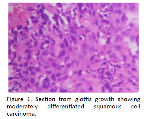
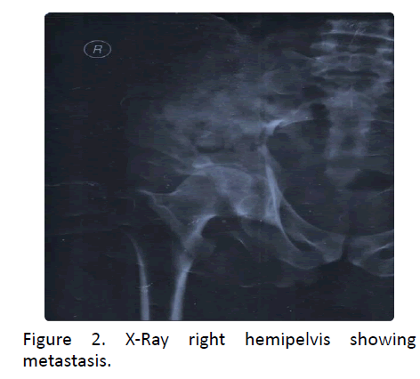
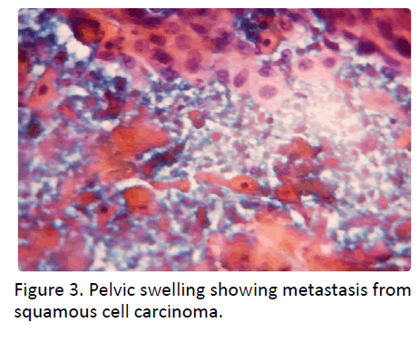
Direct laryngoscopy revealed ulceroproliferative growth replacing left vocal cord, fixed, with congested and edematous left aryepiglottic fold. Biopsy from this growth established moderately differentiated squamous cell carcinoma (Figure 1). Fine needle aspiration cytology (fnac) from left cervical ln was suggestive of metastatic squamous cell carcinoma. X-ray pelvis showed irregular cortical breach with osteolytic destruction involving right superior iliac crest extending to involve entire right iliac bone, lateral margin of acetabulum, adjacent soft tissue appears prominent (Figure 2). Bone scan revealed increased uptake in right superior iliac crest pelvic bone. Fnac from this lesion revealed metastatic squamous cell carcinoma with cystic change and acute inflammation (Figure 3).
Thus the diagnosis of stage iv glottic cancer (t3 n2 m1) with right iliac bone metastasis was established.
After obtaining consent from the patient, palliative radiotherapy 30 gy in 10 fractions to right hemipelvis was planned. After completion of radiotherapy in December 2013 patient achieved symptomatic relief and in view of good general condition was planned with 30 gy/10 fractions to the primary site and neck nodes.
Discussion
The reported frequency of distant metastasis in patients with head and neck squamous cell carcinomas has varied widely, ranging between 6%- 43% in autopsy cases and 8%- 17% in clinical studies [1]. A number of factors have been suggested to influence the development of distant metastasis, such as t classification, n classification, nodal level, primary site of disease, and histological differentiation [2,3]. However glottis lesions rarely develop distant metastasis [4]. Lungs are among the most common sites of distant metastasis. There are no studies published in english literature so far demonstrating pelvic metastasis from primary glottis carcinoma. Present case had advanced disease at presentation with moderately differentiated squamous cell carcinoma with evidence of osteolytic pelvic bone metastasis at initial diagnosis. Present case was treated with local radiotherapy which provided effective palliation.
Conclusion
Thus, a strong possibility of distant metastasis should always be considered in symptomatic patients of advanced head and neck cancer.
References
- Betka J (2001) Distant Metastasesfrom Lip and Oral Cavity Cancer. ORL 63: 217-221.
- Al-Othman MO, Morris CG, Hinerman RW, Amdur RJ, Mendenhall WM (2003) Distant Metastases After Definitive Radiotherapy For Squamous Cell Carcinoma Of The Head And Neck. Head Neck 25: 629-633.
- Papac RJ (1984) Distant Metastases from Head and Neck Cancer. Cancer 53: 342-345.
- Leon X, Quer M, Orus C, Venegas M, Lopez M (2000) Distant Metastases In Head And Neck Cancer Patients Who Achieved Loco- Regional Control. Head Neck 22: 680-686.


