Research Article - Journal of Pathology and Disease Biology (2017) Journal of Pathology and Disease Biology (Special Issue 1-2017)
Pathologic historic mining horses from central Europe.
Cajus G Diedrich*
Department of Biology, Private Research Institute, University of Koblenz-Landau, Ifin, D-56070 Koblenz, Germany
- *Corresponding Author:
- Cajus G. Diedrich
Universitat Koblenz-Landau
Koblenz, D-56070, Germany
E-mail: cdiedri@gmx.net
Accepted on February 10, 2017
Citation: Diedrich CG. Pathologic historic mining horses from central Europe. J Pathol Dis Biol. 2017;1(1):8-33.
Abstract
Early 19th century Deutsche Bund time historic mining horse skeletons were discovered in a mining road water canal nearby the deepest European iron mine west of Prague, Central Bohemia (CZ). Four different sized horse races and interbreeds of larger cold blood horses, medium-sized warm blood Przewalski breeds, but mainly smaller Tarpan breeds such as Ponies cover about six working horse types. Only a single 16 years old female Tarpan breed was buried most complete, such as one more similar old male on the top of it. Other horses were butchered into body pieces, which were fox chew damaged. Most horses died between the ages of 4-7 years due to working stress caused pathologies. The snaffle bit stressed the P2 teeth and caused irregular tooth use, placements or chewing. The thoracolumbar vertebral column was affected in nearly all cases with kissing spine syndrome, articular surface lipping, synovial intervertebral inflamed line/osteoarthritis/intertransverse processes/ankylose osteophytosis as result of permanent heavy wagon pulling. Arthritis is present in several horses’ with synostoses including lipping and nekroses of the tarsals. Very common are synostoses between metacarpi II-IV and between metatarsi II-IV, which seem to result of mining wagon pulling stress, but also of the permanent hard bandage.
Keywords
Deepest iron mine, Historic mining, Graveyard, Taphonomy, Butcher technique, Fox/dog bite marks, Dental, Postcranial pathologies.
Introduction
Beginning of iron mining region Central Bohemia
The Central Bohemian (Czech Republic) region around the city Beroun (Figure 1A) was already explored about 3.500 years ago for Iron in the pre-Roman La Téne Iron Age which people crated larger fortifications such as at Strakonice, west of Prague [1]. Smaller fortifications of the Iron Age period are found east of the village Zdice on the small hill Knihov, and below to the Northern slope a settlement was excavated [2]. The large-scaled mining of the iron ore in this region started somewhere in the 16th century [3]. The 450 millions years old Palaeozoic oolite (pelosiderite) ore rock sediment spreads in length from Úvaly to Plzeň and width from Jince to Zbiroh [4]. The oolite crops out on the surface north of the village Zdice and dips underground in a depth of almost 700 m [5]. The roasted ore rock contains 34-37% of iron [4]. In three mines of the Zdice mining area, from west to east the mines Knižkovice, Hrouda and Černín, about 3 Million tons of the ore rock was taken in the past, which material was moved first by horses [3]. During more than 300 hundred years of mining in the Beroun region of Central Bohemia, horses transported the ore and charcoal of which was burnt at many places on the elongated woody hill ridge north of Zdice.

Figure 1. A. Topography of the Hrouda and Knižkovice iron mines north of Zdice, Central Bohemia (CZ). B and C. Geology of the horse “graveyard” site Zdice, CZ. B. The sediments of the water channel east besides the historical mining road reaching a depth of 2 meters are built of several sand-silt sequences of fluvial origin. C. The darker gravels (with reworked Neolithic/Late Bronze Age shards) and lighter silts allow mapping the boundary of the channel. The main skeletal remains were found on the bottom of the channel within the gravels in depths between 1.5 and 2 m.
The iron ore mines of zdice and its horses in historical reports
At the beginning of the 19th century, extensive iron ore mining begun first in the mine Hrouda northwest of the village Zdice (Figure 1A). The first quarry and other reminders such as old Schacht entrances and collapses are still preserved at the south-western slope of the Čihadlo Hill. The mine Hrouda belonged to the deepest iron-ore mines in central Europe with a mining entrance elevation of 363 m as.l. [3]. In 31 levels and total depth of 760 m for more than a century was mined [3]. In the Hrouda mine was a 600 m long trail where the ore was transported in the mine underground only with the help of “horses” [3]. “Horses” were mentioned to have worked in depths down to the lowermost level and transported wagons with the ore rock between the first and second shaft [3]. Already written is that the mining horses stayed in the mine and lived permanently in the dark with only poor light of the miner’s lamps [3]. Only from time to time, they were transported on the surface [3]. What happened with deathly ill or dead mining horses remained unknown from historical reports of the Zdice mines. Miners used horse-drawn wooden wagons to transport the rock material within the mine, but also for transport to the iron works to the nearby village Knižkovice, in lesser amount to the village Obecnice and later to the closer village Králův Dvůr [3]. For this work, other larger working horse races were used, as it can be demonstrated in this contribution on the excavated horse skeletons from Zdice. The end of the horse use for the transport to the iron work industry was made by the Prague Iron Society which installed in 1914 a modern rope-way from Zdice mine to the Králův Dvůr iron work buildings [3]. The end of the hard underground life for horses came then after World War II, when horses were replaced in the mine with the electrical locomotive [3]. Some buildings are left from the peak of the mining, when a first deep Schacht was opened in 1928/1929, which buildings still exist. The mine was closed already 1962 within Socialistic times [3].
The herein in 2015 rescue-excavated horse skeletons nearby both mines, Knižkovice and Hrouda (Figure 1A) are discussed herein to be either those which worked underground, or which pulled the wagons outside to the iron work factories. Several horse races, their skeleton taphonomy, such as pathologies are used to understand the hard life of the mining/working horses of iron miners in the Central Bohemian region. Although there are few modern compiled reports including historical publications [6,7], there is a gap of knowledge concerning the horse races and their breeding history since 1800, because modern DNA studies reach back in most breeds only the beginning of the 20th century [8]. The herein well preserved articulated skeletons with complete skulls and longbones allow a detail discussion about the different early 19th century mining and working horse races and their possible breed origin. Furthermore, the mining horse graveyard is the first known in Europe, which allows demonstrating the treatment of the “best friends of the mines” after their death. The abundant horse pathologies can be added to historical known illnesses, which are generally not osteological based described. In historic 19th century times, pathologies at horse bones were not be possible to be X-rayed, nor were they studied especially on “working horses” [9].
Material and Methods
The excavated mining horse graveyard area (coordinates: Lat: 49°54'17.80"N, Long: 13°57'37.41"E, after Google Earth) is southwest of the small modern gas station, which was rebuilt in 2014-2016 for the new electric station.
The total amount of 10 skulls (8 with lower jaws, and one isolated lower jaw) from 13 individual horse skeletons/ skeleton parts were found in two cases with nearly complete articulated skeletons, but most horse remains are only “body pieces” (Figure 2A and 2B; Tables 1 and 2). Those were saved within a three weeks rescue campaign as result of a rapid road construction at the end of 2015. Local archaeologists recognized “large amounts” of bones, but they let destroyed skeletons by working with backhoe’s support or left skeletal remains even in-situ, because those were not seen in archaeological (Neolithic/ Late Bronze Age) context (=non-archaeological finds). It was first started to collect some of the visible surface material along the eastern slope. After prospections trenches along the eastern side of the road slope, a systematic larger trench besides the nearly finished new road was made (Figure 3). After success of the first meters, the trench was elongated to the North two times up to 12 m in total length and 1.5-2 m in width (in the main pit area even wider, Figure 3). In both directions no further horse remains were discovered about on to two meters far from the last bone skeleton parts. At least, a part of the graveyard was rescued, whereas many finds along the eastern slope in the further northern part indicate its extension of about 50 m in total length. Only a part of the today mostly destroyed graveyard was saved.

Figure 2. A. Rescue saved articulated horse skeleton parts from the Zdice horse “mining horse graveyard” within a drainage canal which was east parallel to the former mining trail. B. Rescue saved articulated horse parts from the Zdice horse “mining horse graveyard” within a drainage canal which was east parallel to the former mining trail (Photo Diedrich 2015). B. The most complete skeleton 8 in finding position reconstruction, which is the only female (16 years old) and a small wild horse breed of Equus ferus ferus, possibly a Konik horse race.

Figure 3. Taphonomic situation of the mining/working horses from the graveyard within a water drainage channel and pit at Zdice, Central Bohemia (CZ). Data (age classes, sex ): Tables 1 and 2, and details in Figures 4-8. A. Map, B. Photo of Sk 1, C. Photo of Sk 11 skull with neck vertebral column, D. Photo of upper part with Sk 3-7.
| Skeleton | Race | Site | Represented material |
Age | Sex | Butcher signs |
Carnivore bites |
Pathologies | |
|---|---|---|---|---|---|---|---|---|---|
| 1 | Equus ferus caballus breed, large domestic cold blood | Channel (see map Figure 3) |
Skull with mandible (Figure 5), vertebral column (T2-3 absent), pelvis, left femur and distal tibia, astragalus and naviculare | 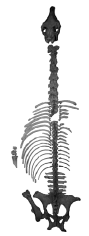 |
4.5 | Male | Both fore and left hind legs are missing, also T2-3, distal tibia with hit cut, thorax hit cut on the right side | For bite damage around pelvis (ischium/ilium) margins | ` Caries on the upper P and M, both LJ P3 to high erupted., Synovial intervertebral osteoarthritis between T15-17 |
| 2 | Equus ferus ferus breed | Channel (see map Figure 3) |
Some middle thoracic vertebrae and costae | No skeleton | 2-3 | ? | No signs | No signs | None |
| 3 | Equus ferus ferus breed, small wild horse breed (=?Konik) | Channel (see map Figure 3) |
Pelvis and last two L5/6 |  |
6 | Male | Hit cut between L5/6 | Few fox bite damage around the pelvis | Right ischium with posterolateral trauma, synostotic L5/6, Synovial intertransverse processes of L5/6 |
| 4 | Equus ferus caballus breed, large domestic cold blood | Channel (see map Figure 3) |
Mandible, vertebral column (T10-L6) and sacrum, posterior ribs, left and right fore leg (ulna/radius, carpalia, mc I-IV, ph I-2) | 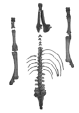 |
4.5 | Male | Thorax hit cut on the left side | Fox bite damage on the vertebrae spines | Synostotic metacarpalia on both sides |
| 5 | Equus ferus ferus breed, small wild horse breed (=?Konik) | Channel (see map Figure 3) |
Pelvis, T18-L6, right femur and half proximal tibia | 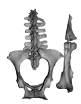 |
16 | Male | No signs | Fox bite damage on the vertebrae spines and around pelvis (ischium/ilium) margins | Coxofemoral osteoarthritis, Vertebral ankylose osteophytosis in combination with Synovial intervertebral osteoarthritis, and Intertransverse processes at the L5/6 |
| 6 | Equus ferus przewalskii breed, medium-sized wild horse and (=?Konik) interbreed | Channel (see map Figure 3) |
Skull with mandible (Figure 5), vertebral column (C4-T6 absent), nearly all left costae, rigtbhn costae half, pelvis, left tibia to ph III, left fore leg without scapula, right radius, carpalia and half Mc III | 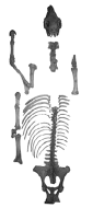 |
4.5-5 | Male | Thorax hit cut on the right side, hit crack damage of right radius and Mc III | Fox bite damage on the vertebrae spines and around pelvis (ischium/ilium) margins | Mc II-IV synostotic, carpalia with exostoses, Fore- and Hind leg osteoarthrosis (including nekrosis, exostosis, lipping), Articular surface lipping with syndesmophyte at L5/6 |
| 7 | Equus ferus ferus breed, small wild horse (=?Konik) with pony interbreed | Channel (see map Figure 3) |
Skull with mandible and stylohyoidea, incomplete fore legs, T7-L6, pelvis, no hind legs | 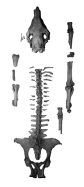 |
6 | Male | Vertebral column cut (T16/17), thorax cut on both sides, right and left fore leg bones hit damaged at several places, right Mt III hit cracked | Fox bite damage on the vertebrae spines and around pelvis (ischium/ilium) margins | Incisive teeth polished anterior incorrectly |
| 8 | Equus ferus ferus breed, small wild horse breed (=?Konik) | Pit (see maps Figures 3 and 7) |
Nearly complete and articulated skeleton | 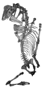 |
16 | Female | Thorax cut (damaged middle costae) on left side | Fox bite damage on both humeri proximal joints, and around pelvis (ischium/ilium) margins | P2 to much uplifted and with convex polish on anterior side, complete tooth rows to much concave/convex, upper P/M with caries, . Intertransverse processes at the L5/6, synostoses on all Mc, Fore- and Hind leg osteoarthrosis (including nekrosis, exostosis, lipping), Articular (spat) |
| 9 | Equus ferus ferus breed, small wild horse breed (=?Konik) | Pit (see maps Figures 3 and 7; found above Sk 8) |
Nearly complete and articulated skeleton | 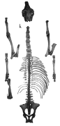 |
16 | Male | Thorax cut (damaged middle costae) on left side, left humerus with hit mark | Bite damaged along distal margins of the ilium/ischium, and proximal humerus joints chewed | Right I2 broken and shorter, both M1 caries, right I1-2 moved and I3 broken |
| 10 | Equus ferus ferus/przewalskii breed, medium-sized Przewalski and (?Konik) interbreed | Channel/pit margin (see map Figure 3) |
Skull and probably to this belonging scattered parts of the skeleton |  |
16 | Male | Hit damage of brain case | Intertransverse processes at the L5/6 and posterioventral (articulation with sacrum) disc laison with dystrophic mineralization and ventral herniation at L6 | |
| 11 | Equus ferus ferus breed, small wild horse breed (=?Konik) | Channel (see map Figure 3), in two axial parts and leg, and part of rib cage |
Skull with | 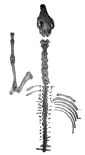 |
5.5 | Male | Axe cut between C7/T1 and again posterior at L2, thorax cuts | dispersed in three body parts | All metacarpi on fore leg synostotic, Synovial intervertebral inflamed line |
| 12 | Equus ferus ferus breed, small wild horse breed (=?Konik) | Channel (see map Figure 3) |
Skull with complete vertebral column and nearly complete thorax such as pelvis and left femur such as its proximal tibia |  |
4.5 | Male | Axe cut on the upper third of the left tibia | Bite damaged along distal margins of the pelvis (ilium, ischium); bite marks on the distal nasals | Irregular I3 change, between T11-L6, Articular surface lipping between posterior T and L2 |
| 13 | Equus ferus caballus, Pony breed | Channel/Pit margin (see map Figure 3) |
Pelvis, right femur, and right incomplete fore leg | 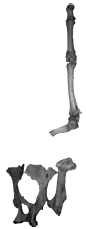 |
6 | Male | No signs | Bite damaged on proximal humerus (chewed of completely), and on distal Ph II | Exostoses laterally on Trochanter major |
Table 1. Main data to the mining/working horse skeletons from the graveyard of the Zdice iron ore Mines, Central Bohemia (CZ).
| Skeleton | Skull | Mandible | Humerus | Radius | Mc III | Pelvis | Femur | Tibia | Mt III |
|---|---|---|---|---|---|---|---|---|---|
| 1 | L=650, W=216, Tooth row P2-M3=175 |
L=495, H (P4/M1)=77, Tooth row P2-M3= 177 |
- | - | - | W=620, AcetW=73 |
L=460, DW=110 |
DW=87 | - |
| 2 | - | - | - | - | - | - | - | - | - |
| 3 | - | - | - | - | - | L=440, W=521, AcetW=66 |
- | - | - |
| 4 | - | L=450, H (P4/M1)=90, Tooth row P2-M3=200 |
- | L=405, DW=89 |
L=256, DW=47 |
- | - | - | - |
| 5 | - | - | - | - | - | AcetW=67 | - | - | - |
| 6 | L=580, W=214, Tooth row P2-M3=187 |
L=445, H (P4/M1)=86, Tooth row P2-M3=187 |
L=315, DW=85 |
L=380, DW=85 |
L=237, DW=57 |
L=460, AcetW=73 |
- | L=396, DW=83 |
L=285, DW=56 |
| 7 | L=545, W=201, Tooth row P2-M3=173 |
L=425, H (P4/M1)=66, Tooth row P2-M3=174 |
DW=88 | - | - | W=518, AcetW=69 |
- | - | |
| 8 | L=570, W=216, Tooth row P2-M3=146 (without P1) |
L=432, H (P4/M1)=75, Tooth row P2-M3=175 |
L=311, DW=86 |
L=355, DW=84 |
L=238, DW=52 |
- | L=410, DW=97 |
L=390, DW=83 |
L=280, DW=52 |
| 9 | L=562, W=205, Tooth row P2-M3=173 |
L=447, H (P4/M1)=65, Tooth row P2-M3=173 |
DW=85 | L=381, DW=81 |
L=246, DW=52 |
- | DW=84 | - | L=285, DW=53 |
| 10 | W=210, Tooth row P2-M3=162 (pathological) |
L=453, H (P4/M1)=80, Tooth row P2-M3=175 |
- | - | - | L=442, W=465, AcetW=65 |
- | - | - |
| 11 | L=565, W=200, Tooth row P2-M3: 182 |
L=435, H (P4/M1)=77, Tooth row P2-M3=182 |
L=305, DW=80 |
L=350, DW=75 |
L=230, DW=50 |
L=485, W=550, AcetW=70 |
L=425, DW=98 |
- | - |
| 12 | L=532, W=207, Tooth row P2-M3=165 |
L=415, H (P4/M1)=75, Tooth row P2-M3=160 |
- | - | - | L=400, DW=97 |
- | - | |
| 13 | - | - | L=295, DW=75 |
L=329, DW=73 |
L=216, DW=48 |
AcetW=65 | L=395, DW=90 |
- | - |
| Skull 1 | L=558, W=214, Tooth row P2-M3=168 |
- | - | - | - | - | - | - | - |
| Skull 2 | L=560, W=210, Tooth row P2-M3=167 (with P1) |
- | - | - | - | - | - | - | - |
Table 2. Main osteometric data of the skeletons from the mining horse graveyard of the iron ore Mines, Zdice, Central Bohemia (CZ), all in mm: H (P4/M1=Height of mandible behind P4, W=Skull width between frontalia, L=Length, DW=Distal joint width.
There are three “concentrations” within and along the channel which were mapped for taphonomic studies (Figure 3). Those allowed distinguishing: 1. Articulated individual skeletons, skeleton parts, leg or axial skeleton parts, or single bones, and 2. Anthropogenic hit marks versus carnivore bite damages. After washing and gluing of the fragile decalcified and in most cases modern broken bones, the details of the pathologies and hit/cut/ bite marks became well visible.
The horse bone material was determined using different anatomy publication sources [10-14]. The horse age classes are estimated by: 1. Tooth replacement stage [15,16] and 2. Bone fusion degrees (especially vertebra disc fusion) and pathologies (Figure 4). Those were essential to understand spread or partly dismembered skeleton parts to originate from one individual each. Dental and postcranial pathologies were compared with archaeological, historical and modern descriptions of horses [9,17-21]. The sex is obvious to determine on the skulls and lower jaws by the canine presence in males (Figure 4) [9]. Finally, the body sizes and heights were not calculated [22,23], instead skull metrics and longbone (humerus, radius, metacarpus III, femur, tibia and metatarsus III; Figure 5) metrics according to Müller [22] and May [24] were compared between the races directly.

Figure 4. Main skull/mandible characters and shapes (mandible and cranium heights between P4/M1) of the Zdice mining graveyard horses and correlation to modern skulls of domesticated and rebreeds wild horses and Late Pleistocene wild horse (from different collections) and head profile shapes (head types modified from Kapitzke, 1993). Most probably, the smaller Zdice horses originate from Polish Konik (Tarpan breed) horse breeds (e.g., female Sk 8 and male Sk 9 skeletons). The Konik breed seem to have been crossed with a pony. Furthermore a Przewalski breed is represented in Zdice which must have been crossed with a Konik. Those smaller-medium and small sized horses were the underground mining horses. The largest horse race is a warm blood, a working horse of unclear race for pulling wagons outside the mines.

Figure 5. Comparison of the age classes on complete skulls (tooth use method) and postcranial longbones (joint fusion method) of the mining horses from Zdice.
Modern horse skulls and some longbone material were used for a direct and in some cases for a metric comparison from the following collections: Naturhistorisches Museum Mainz, Germany (Przewalski horse skull), Julius-Kühn-Museum der Universität Halle-Saale, Germany: Przewalski horse skeleton), and private collection F. Menger, Germany (Pleistocene Tarpan skull). In this contribution only three main cranial metrics (skull: length, tooth row lengths and nasal width; longbone length and distal width) were taken to distinguish the size classes and races (Figure 5). More useful for race relationships and breeding questions (domestic and wild horses) are herein selected skull and mandible characters. The different mining horse skulls, a Late Pleistocene Tarpan horse skull and modern caballoid horse skulls were figured (Figure 6) to compare their lateral profiles with the main head form shapes and race types of modern races sensu Nissen [25-29], and including newer DNA study results on caballoid (=domestic) and wild horses [8,30]. E. g. important is the skull shape similarity of Przewalski horses and Zebras, which are DNA tested very close to each other [8] sharing therefore similar osteomorphologic characters (e.g., lower jaw shape). Singular metric metapod studies do not allow further successful detail discussions about the races, as being demonstrated herein on the skull material and correlation to the metapod sizes.

Figure 6. Comparison of the four main-sized mining horses in their postcranial longbone metrics from Zdice. In some cases longbones are not present and the metrics was used instead of modern similar sized horses (especially in caballoid breeds of cold blood and pony).
Results and Discussion
The age of the horse skeletons
The “horse graveyard” bone material is after historical descriptions [3] dated coarsely between 1806 and 1914. The site is directly on the historical road which connected already at the beginning of the mining (beginning 19th century) the Hrouda and Knižkovice mines (Figure 1A) [3]. A half, only inside yellow-green glazed henkel pot fragment found above the horse skeleton layer, is typical for pottery around 1800 [31,32]. The excavated horses are therefore expected to be from the first half of the 19th century. It seems, the older horse skeletons were recovered further from the older road dug oval pit, and the younger ones are from the water drainage channel (Figure 3).
Geology
The fluvial sediments of the horse skeleton bearing fine gravel/ silt layers (Figure 1B) cannot originate from the nearby smaller streams which are too far (no high flood impact) and in too low elevations (about 5 meters below today’s stream elevation). The only explanation of their presence is a water drainage channel (Figure 1C) which reached about 2-2.5 m deep east besides the historical road for dewatering reasons of the Knižkovice village. The already deepened U-shaped trail (about 1.5-2.5 m) resulted from strong use by heavy wagons and horses. It seems to have been refilled on its margin, especially east side with erosive sediment and changing by humans deposited layers of burnt charcoal (above the bone layers). Such burnt material is not present in the section (Figure 1C) nor was it used to cover the horse skeletons.
The Zdice mining horse races and interbreeds
First, the complete mining horse skulls only of males from Zdice were used to compare (Table 1), whereas no metric analyses were made. Such studies are initially started, but not satisfying useful for a good separation, whereas some characters let distinguish skull shape types [33,34]. The direct comparison show very obvious different useful characters already in their lateral profile (Figure 6): 1. Skull length/nasal width and tooth row length sizes. The three main metrics (Table 2) let distinguish three size-classes on the historical material, but a fourth including a modern Pony skull (because pony is present with postcranial material). 2. Nasal shape: a. Straight (plesiomorph character in Tarpan wild horses), b. Convex (plesiomorph character in Przewalski wild horses), c. Convex (=domestic character in caballoid “cold blood” horses). 3. Lower jaw shapethere is a strong difference of the jaw shape, whereas the metrics used herein to distinguish those was measured for the mandible height between the P4/M1 teeth (Figure 6; Table 2): a. Nearly straight basal mandible with medium heights, b. Convex profile of the mandible (typical in Przewalski horses and to those by DNA tested close related Zebras), c. Low mandible height (typical for Tarpan horses), and d. Very low mandible height (typical for Ponies). The mandible shape (plus skull characters) allow finally also speculating on interbreeds between the four main race types (Figure 6). 4. Presence and absence of the P1 (=“wolf tooth”)-a. Presence (typical in Pleistocene Tarpan wild and rare in Przewalski horses (or caballoid horses) and therefore a plesiomorph character), b. Absence (rarely present and generally absent in domestic horses). The four main shape skull types from Zdice were compared to the head profiles of four modern horse breeds, which allow their classification first in four race types, and possibly three interbreeds (Figure 6).
Large-sized caballoid domestic cold blood-The large horse skull (Sk 1), being represented by two skeletons (Tables 1 and 2), has compared to the other types from Zdice: a. Convex nasal area, b. Long distance between P2 and I3, c. Medium mandible height between the P4/M1, and is d. The largest form (650 mm in total cranial length). Compared to the head profiles illustrated by [29], this fits to a large cold blood domestic working horse shape called “Ramshead”, such as typical for modern Münsterland, Belgiens [29], or Czech cold blood breeds, which latter was breed not before the beginning of the 20th century [35], and can therefore not represent the same race as found in Zdice.
Medium-sized Przewalskii horse breed- The medium-sized horse skull type is represented by one skull (Sk 6, Figure 6), the other one (interbreed) has a brain case hit damage. The direct comparison with a modern Przewalski horse skull (also representing already rebreeds [36]) fits exactly in its shape and size and its wild horse characters mentioned above, which are: a. Concave nasal area, b. Short distance between P2 and I3, c. Large mandible height between the P4/M1, and is, d. The medium-sized form (580 mm in total cranial length, Table 2). The head profile fits to a medium-sized wild horse shape called “Ramsnose”, being typical for Przewalski horses and interbreeds [29,36].
Small-sized Tarpan horse breed- The smaller horse skull type from Zdice is represented by four complete skulls and two crania including both, males and females (Sk 7-9, Figures 3 and 5). The direct comparison to a wild horse Tarpan skull from the Late Pleistocene Glacial fits exactly in the shape of its typical slender mandible, whereas the cranium shares the characters of the above Przewalski horse, which Tarpan skull characters are: a. Concave nasal area, b. Short distance between P2 and I3, c. Low mandible height between the P4/M1, and d. Small-sized form (550 mm in total cranial length). Additionally, only this oldest wild horse race breed has in two skulls the upper jaw wolf teeth (P1) teeth (Figure 12A), which is a typical character for “ancient horse types” [29]. The head profile fits a horse type shape called “Straighthead” (Figure 3) [29]. However, interesting is the fact, that primitive domestic breeds with plesiomorph characters from Tarpans around 1780 are mentioned as Konik Horse from Poland [8,28,29,37]. Most probably, such Polish Konik domestic Tarpan races [38] were used as mining horses in Zdice or Czech Republic mines in general, which also would correlate to their domestication time at the end of the 18th century. The complete Tarpan breed skeletons of 16 years old male/female as oldest burials of Zdice (pit area Figure 3) date to the beginning of the 19th century and might come from the polish Konik “population”.
Smallest-sized Pony horse-The smallest horse type skull is sadly not present, but it is represented with parts of the postcranial skeleton (Figure 5, Tables 1 and 2). Herein, a modern Pony skull is figured to demonstrate its similarities in some of the Konik/Tarpan breed skull mandibles, which are either more “wild horse –like” or much slimmer such as found in Ponies. The skull shape of the Pony is close to the “Wedgehead” (Figure 6) [29].
Interbreeds-Using the herein described characters, there seem to be three interbreed races (Figure 6): 1. Cold Blood /Przewalski interbreed, because the mandible and postcranial leg bones have very large sizes, but the mandible has Przewalski horse characters, 2. Przewalski/Konik=Tarpan breed, because of their mixed characters in the skull and mandible, 3. Konik=Tarpan breed/Pony, because of the very characteristic shaped mandible, but skull sizes and shapes of the wild horse.
Graveyard horse skeleton taphonomy
There are two main horse deposits along the road which were made in different times. First, a large oval pit was dug, into which the two complete female (Sk 8) and male (Sk 9) horses were dropped on each others as a whole (Figure 3). All other skeleton parts obviously are from a younger period, when the channel was cutting the older oval horse burial pit. Their time of deposition each can not be dated in more detail. Important are those taphonomic and time differences between the pit (complete and possibly breeding horses of smaller Konik horse race types) and channel (ponies, przewalski horses and large working horses mainly in body parts).
Compared to articulated skeletons from other graveyards or even horse graves in the archaeological record of the European Neolithic [39], Orient Byzantines [24], European Merovingian, Saxonian and Slawic-Awarian [23,40,41], Skandinavian Vikings [42], European Medieval [43,44], and different aged archaeological sites in the Czech Republic [45], the historical mining horse graveyard of Zdice is very different compared all. Unique are the position of the graveyard along a road (water channel) and further from the mine or settlement, and the butchering of the horses into similar body parts to fit into the water canal, or c. Presence of selected strong pathologic/ill horses. Furthermore, the thorax openings on one or two sides (Figures 7-9) are also not reported in the above mentioned publications, such as are hit-cut and hit broken bone damages:

Figure 7. Taphonomic situation–selected examples (positions see Figure 3). Stage 1: Only the female 16 years old horse is nearly complete, but has as the others the lateral thorax rib cage opening=all ribs are cut in the upper third. This cut most probably by an axe also caused the leg damages (hits). In skeletons 8 and 9 chewing of a fox can be observed on the pelvis or all proximal joints of the humeri. Stage 2: In skeleton 9 the hind legs were removed, also fore legs, but those were dropped besides (without shoulder blades). Similar it is in skeleton 6, but more disarticulated and clearly in the shoulder area cut fore legs as result of the right thorax cut (therefore left leg is “complete”). Stage 3: All legs are missing in skeleton 3, which consist of the skull, mandible, nearly complete vertebral column (two anterior T are missing due to removal of fore legs in the shoulder area) and pelvis, whereas a distal large tibia and tarsalia demonstrate with a diagonal hit cut the leg removal by large iron tools (see axe fits in Figure 8).

Figure 8. Taphonomic situation–selected examples of arranged skeletons 1, 9 and 11 (Figures 3 and 9). One side (Sk 1/9) and two side (Sk 11) thorax cut openings. Whereas the ribs and vertebral column was cut by a large metal tool (see below axe fits, =hit cut), the legs were mostly hit (=hit crack) with the opposite non-cutting side of the axe (details Figure 9).

Figure 9. Taphonomic situation–hit and cut damaged leg longbones and axial skeleton vertebrae at the mining horses from Zdice.
Hit/cut damages-Two types of anthropogenic caused bone damages are figured with good examples (Figure 9). Hit damage and spiral breakage is present in nearly all cases on the fore and hind legs. The positions are found in the middle shaft of the humerus (e.g., hit hole Sk 9, Figure 9), in the upper middle and lower part of the radius (which caused also the flaking of the fused ulna), and in the upper and middle part of the metacarpus III (which caused also the flaking of the neighbored Mc II and IV). In Sk 6 the fore leg has at minimum two hit cracks in the middle of the radius, and the Mc III, which were found finally in three parts scattered (Figure 3). The hind leg hits were made in the upper, middle or distal part of the tibia, whereas only in one case (Sk 12) the left hind leg was cut below the tibia joint (articulated pelvis, femur and proximal tibia–Figure 3, Table 2). Furthermore, hits are found also on the Mt III. Rarer are clear signs of butchering of the axial skeleton. The best example is Sk 11, which was separated in two column parts (Figure 3, Table 2) with a clear cut mark on both; the C7 and T1 (Figures 8 and 9). Both column parts were found even some meters far from each other (Figure 3). This cut was made between neck/shoulder blades. Another main cut area is found between the T16 and L6 to separate the posterior body part. Other examples are found in Sk 3, 7, 11 (Figures 8 and 9). Cut marks are rare on all skeletons, and only one good example at a thoracic rib was found at Sk 8, which furthermore demonstrates a non-sharp cutting edge of a large metal tool (axe/adze) that left several parallel with 90° angle to the cut striations (Figure 9). The absence of small metal tool cut marks fit to a non-filleting or non-use of the horse meat as food for the miners or their dogs.
Carnivore impact- All signs of carnivore bite damage (Figure 10) are found after the butchering, which is obvious by the overlap in some cases of bites at hit cracked/cut bones and bite marks. In most cases, those are found at the proximal humerus joint, which is in many cases half to completely chewed of, even including the complete Sk 8 (Figures 7 and 10; Table 2). This is of importance because the scapula was cut in nearly all cases from the humerus (with the thorax cut–Figures 7 and 8) damaging in some cases the proximal humerus joint (proximal part chopped off). There is one example outside the mapped area of such a cut proximal humerus joint cap. The problem to prove this in most other humeri is the overprint in all incomplete ones by the bite chew damage (Figure 10; Table 2). In several cases (e.g., Sk 8, 9) the spongiosa was even scratched deeply leaving a larger cavity always on the outer lateral joint area. Such similar dog bite damages are also described from archaeological site horses [46]. This was more easy and possible, if the upper part of the humerus joint was already cut. The carnivore impact can not explain the absence of the scapulae in all incomplete skeletons. Only in the complete skeleton 8, those are still in articulation, but whereas the humeri joints are again with larger cavities, the scapulae are intact in their proximal joints. Scapulae were clearly not removed from the carcasses by carnivores (dog/fox) and are suggestive of a human butcher context. Similar is the absence of at least one femur in articulated skeletons (Table 2). Furthermore, bite damage is found along the distal spines of several vertebral columns strongest in the area of the anterior thoracic vertebrae (e.g., Sk 8). The bite marks are found at the pelvis, especially with Zig-Zag margins (Figure 10). There are generally two sizes of bite scratches at least: 1. 4-5 mm wide scratches (only one humerus joint, Sk 8, Figure 10), 2. 2-3 mm wide scratch marks, or impact marks of I, C and P or M teeth. The most important bite impacts are found on the inner side of the horse carcass–the ventral side of thoracic to lumbar vertebrae (Figure 10). At three examples (e.g., Sk 11) the distances of parallel canine bites are available to measure: 20 and 24 mm distances (Figure 10). Finally, incisive impact marks are present with best as example at the ileum margin of Sk 7 (Figure 10). Important is the space in-between those incisive bite holes, which allows for the identification of the carnivore.

Figure 10. Carnivore dog/fox bite marks and bone damage examples on the mining horses from Zdice, with bite fitting experiments using a modern fox skull. A. Larger bite scratches and cavity in the spongiosa of the humerus proximal joint of Sk 8. B-D. Fox bite marks left in the ventral side of Sk 11 thoracic vertebra centra or distal spines. E and F. Zig-zag margins resulting from the breaking scissor teeth are found on the lateral margins of pubis or ischium of most of the horse skeletons (Table 1).
The bite scratch width, and bite distances of incisici/canini allow identifying two different sized carnivores. Compared to bite damages on prey produced by dogs (Canis lupus familiaris), only in one case (humerus) their short presence can be proven. In nearly all other cases, the bite sizes fit more to maximum fox sized predators. A modern Vulpes vulpes skull and lower jaw fit exactly into bite marks on a pelvis and vertebrae with the upper (24 mm) and lower jaw (20 mm) in their canine distances (Figure 10). Refitting of the bites into the bite marks are even more convincing using the anterior incisives, which I1-3 is in the fox skull visible in about 1 mm in distances which identifies the main horse scavenger as the red fox. Finally, this small scavenger is also represented from the graveyard with one incomplete humerus. Their “permanent” presence at or nearby the site is important to understand the taphonomy of the horse carcasses, and the question about a possible covering with sediment after their dropping into the pit/channel. As listed in Tab. 1, nearly all horse skeletons/skeleton parts have fox scavenging damage marks, therefore the horse body parts seem to have been not covered by soil at times. Most probably, the horse carcasses were even thorax cut to get easier access to the intestines/inner organs to feed by foxes. Those scavengers seem to have not removed any heavy body parts, but played a role in the dismembering of the one or other carcass remains, but only in short-distance. Furthermore, dislocation must have happened to some bones by stronger fluvial impact (e.g., storm rain events) within the water drainage channel.
Pathologies
The cranial dental and postcranial axial and leg pathologies listed for each horse skeleton (Table 1) are figured in most cases (Figures 11-17). The position of pathologies (together with butcher areas) on the Zdice horses is compiled in Figure 18.

Figure 11. Dental pathologies 1–A. Sk 10 skull with strongest dental pathologies at all teeth, ventral view. B. Labial view of the dentition, C. Dorsal and labial views of the lower jaw dentition, D. Cranial view of the incisive dentition, E. Sk 7, cranial view of the incisive dentition, F. Sk 9, cranial view of the incisive dentition.

Figure 12. Dental pathologies 2–A. Sk 8, occlusal and labial views of the upper and lower jaw dentition, B. Sk 4, occlusal and labial views of the lower jaw dentition.

Figure 13. Postcranial pathologies vertebrae–A and B. Synovial intervertebral inflamed line and C. Articular surface lipping at younger individual Sk 12 (4.5-6 years) between T15 and L2. D and E. Articular surface lipping at Sk 7. A. Lateral left, B. Detail, C. Cranial, D. Cranial, E. Lateral right, F. Cranial.

Figure 14. Postcranial pathologies vertebrae–A-B. Articular surface lipping with syndesmophyte at Sk 7 L5/6. C and D. Intertransverse processes at the L5/6 and posterioventral (articulation with sacrum) disc laison with dystrophic mineralization and ventral herniation at L6 of Sk 10. E and F. Synovial intervertebral osteoarthritis (or “bamboo spine”) between T15-17 at Sk 1. A. Caudal, B. Ventral, C. Caudal, D. Dorsal, E. Lateral left, F. Dorsal.

Figure 15. Postcranial pathologies vertebrae–A-E. Vertebral ankylose osteophytosis in combination with Synovial intervertebral osteoarthritis (or “bamboo spines”), and Intertransverse processes at the L5/6 at Sk 5. A. Dorsal, B. Lateral left, C. Ventral, D and E. Details of the ventral view (syndesmophytes).

Figure 16. Postcranial pathologies pelvis/hip joint–A-C. Trauma at the right ischium. D. Exostoses at the Trochanter mayor of the pony skeleton Sk 13. D-G. Coxofemoral osteoarthritis in Sk 5. A. Lateral view, B. Caudal view, C. Dorsal view. D. View on joint, E. Lateral, F. Ventral, H. Lateral.

Figure 17. Postcranial pathologies legs–A, F-K. Fore-and Hind leg osteoarthrosis (including nekrosis, exostosis, lipping) at Sk 6 and 8. B-E. Metapodial synostoses at Sk 4, 6, 8, 11. A, F. Cranial, B-E. Lateral, C. Caudal, all other different views on the joint surfaces.

Figure 18. Bone terminology used herein, pathology and butcher technique overview at the mining/working horses of the graveyard site Zdice, Central Bohemia, CZ.
Dental pathologies–were compared to the works of Launer et al. own observations in horse stables. At some of the Zdice horse skulls upper P/M teeth caries holes are visible in the dentin (Sk 8, 10, Figures 11A and 12A). Nearly none of the skulls have normal tooth rows, or straight occlusal tooth row surfaces, also the jaw joints are mostly non-smooth and irregular in shape and their joint convexity. Generally, in all dentitions the first lower jaw P2 is too much uplifted, and has in some cases concave abrasion (Figures 11B, 12A and 12B