- Biomedical Research (2014) Volume 25, Issue 4
Detection of diabetic retinopathy by applying total variation.
Poornima S V, Parvatha Lakshmi B, Nishchala T K, Umamakeswari ASchool of Computing, SASTRA University, Tanjore, Tamil Nadu, India
- *Corresponding Author:
- Poornima S V
School of Computing, SASTRA University
Tanjore, 613401, Tamil Nadu, India.
Accepted date: August 26 2014
Abstract
The incendiary changes in the retina will lead to curative approaches that prevent blindness. Diabetic retinopathy is the most prevalent diabetic eye disease that produces intensive damage to the retina by creating progressive changes in its blood vessels. This can eventually lead to blindness. Diagnosis of this disease in its earlier phase is very important and this can be accomplished by a comprehensive eye examination and regular screenings. During the screening process, the images of the retina are taken by making use of an ophthalmoscope or a fundus camera. In this work, an attempt has been made to differentiate between an retinopathic eye from an healthy eye by applying total variation to the fundus photographs. Total variation measures the signal variation between successive signal values. Understanding
Keywords
Diabetic retinopathy, screening, ophthalmoscope, fundus images, total variation, HRF dataset
Introduction
Human eye is a fundamental sense organ that enables vision. It is considered a natural camera. The retina is an indispensable part of the human eye that consists of rod and cone cells. Diabetic patients are highly vulnerable to deteriorating diseases such as diabetic retinopathy. It is a complicated disease that generally prevails in patients with chronic diabetes. Diabetic retinopathy at its worst causes complete blindness. There are a few screening methods that can detect early diabetic retinopathy. If detected early, the progression of diabetic retinopathy can be curbed by keeping blood sugar levels under control. Ophthalmoscopy is one of the screening methods that can be used to detect the disease at an early stage. But ophthalmoscopy requires the expertise of skilled professionals. An alternate strategy is to perform fundus photography.
Diabetic retinopathy [4,5] is a chronic disease causing massive damage to the retina, resulting in an alarming damage to human sight due to the destruction of blood vessels. This condition commonly affects both the eyes. There are two categories of diabetic retinopathy.
1) Non-proliferative Diabetic Retinopathy
The non-proliferative kind is the initial stage of diabetic retinopathy that causes leakage of blood and leads to swelling in retina. The blood vessels in the eye swell and result in leakage of lipids and blood that are called hard exudates. These deposits cause swelling along fovea centralis and flattening of the macula, a condition called macular edema resulting in loss of clear central vision. This can be treated using laser surgery [10]. The blood leaked into the vitreous humour often leads to occlusion and could be corrected by vitrectomy.
2) Proliferative Diabetic Retinopathy
Another type of diabetic retinopathy is known as proliferative diabetic retinopathy. The major cause for this disease is uncontrolled diabetics [9]. Nonproliferative diabetic retinopathy progresses into proliferative diabetic retinopathy, if diabetes is not controlled.
As NPDR results in insufficient blood supply to eyes, the system responds to it by developing new blood vessels, a process called neovascularization. These newly developed blood vessels are often weak and are prone to leak. This is called PDR or proliferative Diabetic Retinopathy.
If the new vascular tissues arise on or very close to the optic disk, they are called NVD-PDR or neovascularization on the disk. This type of diabetic retinopathy will finally lead to blindness. The other type of PDR is called NVE-PDR. This disease is marked by the growth of neovascular tissues. The newly grown neovascular tissues will not be very near to the optic disk.
Screening for Diabetic Retinopathy
Ophthalmoscopy [6] is a well-known screening technique for diabetic retinopathy. It must be performed by a professional in an office setting. The inside of the fundus is inspected and studied using an ophthalmoscope. Another well-known screening method is fundus photography. These fundus photographs [7] can recreate areas of the fundus that are larger than those that can be seen using an ophthalmoscope. The present study is carried upon images obtained from fundus photography screening have been used for an automatic detection of diabetic retinopathy.
Material and Methods
An effective screening method must fulfil certain criteria. The sensitivity and the specificity must be high. In other words, the number of true positives detected must be high, while the specificity or the number of true negatives eliminated must be high as well. Even though there are several screening methods to detect diabetic retinopathy, debate prevails on how this screening is done. Ophthalmoscopy allows skilled professionals to look into the inner structure of the eye through an ophthalmoscope. Fundus photography is an alternative for this method. [8] It creates images of inner structure of an eye that are later examined by trained professionals.
The focus of our work was to automate the detection of diabetic retinopathy, thereby eliminating errors culminated by human measurement. Fundus images obtained from HRF database [2] have been used for this study. This database is a public repository containing high-resolution fundus images of retinas of healthy and retinopathic eyes, and thus serves as a benchmark for algorithms aiming at automation. The screening was carried over 100 diabetic subjects of 25-90 years of age and thirty photographs after the screening have been randomly selected and made available as a public repository. This dataset consists of 15 images of healthy eyes and 15 images of retinopathic eyes. The pictures taken by an expert using a Canon CF-UvI fundus camera have a resolution 3504 x 2336.
Total variation is been applied on the images of both healthy and retinopathic eyes. Total Variation [1,3] is a common method used in digital image processing. According to this method, signals that contain spurious information have high amounts of total variation. Two adjacent pixel values are compared and the variation in them is calculated. Colour images are converted to grayscale images before taking them as input since grayscale images make the comparison much easier.
The values obtained on performing total variation on healthy eye images and retinopathic eye images have been compared. Using this method, a healthy eye is differentiated from an retinopathic eye. Since this method has worked out well for these benchmark images, it is expected to work well for other images and thereby help in the detection of diabetic retinopathy.
Results
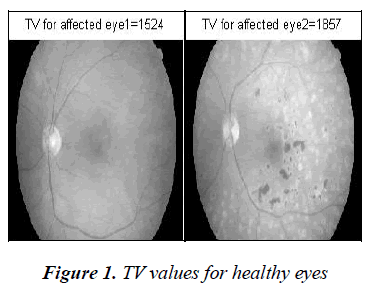
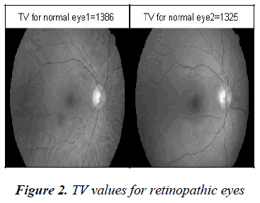
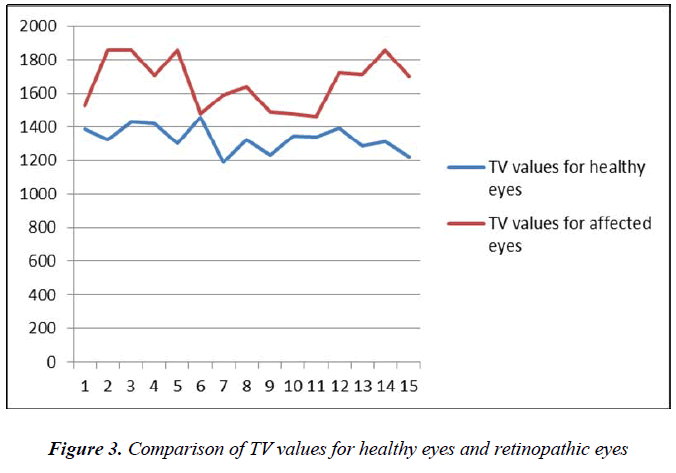
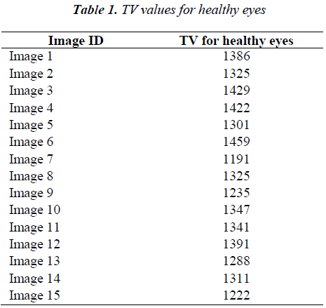
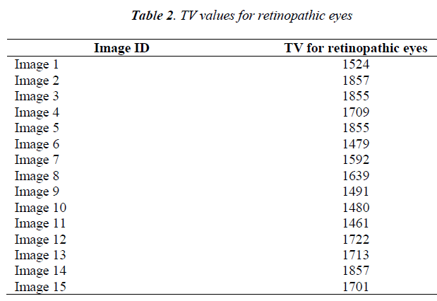
Total variation values for eyes with diabetic retinopathy are significantly high when compared to those of healthy eyes. Table 1 presents the TV values for some healthy eye images and table 2 displays the TV values for retinopathic eye images. Figure 2 and Figure 3 illustrate the TV values for normal eyes and retinopathic eyes.
Discussion
In the present, an attempt to compare the total variation values for healthy eyes and eyes with diabetic retinopathy is made. Total Variation methodology is applied to classify the fundus images as healthy or retinopathic eyes. The images obtained from HRF dataset are used as standard reference. The TV values for retinopathic eyes are significantly high when compared to the TV values for the healthy eyes. The graph (Figure 3) demonstrates that the TV values of healthy eyes and retinopathic eyes do not overlap. So, this method is a good screening strategy for diabetic retinopathy.
References
- Chan TF, Golub GH, Mulet P. A Nonlinear Primal-Dual Method for Total Variation-Based Image Restoration. SIAM Journal on Scientific Computing. 1999 Jan;20(6):1964–1977.
- Kohler T, Budai A, Kraus MF, Odstrcilik J, Michelson G, Hornegger J. Automatic no-reference quality assessment for retinal fundus images using vessel segmentation. IEEE CBMS; 2013 : 95–100.
- Osher S, Burger M, Goldfarb D, Xu J, Yin W. An Iterative Regularization Method for Total Variation-Based Image Restoration. Multiscale Modeling &Simulation. 2005 Jan;4(2):460–489.
- Owens DR, Gibbins RL, Kohner E, Grimshaw GM, Greenwood R, Harding S. Diabetic retinopathy screening. Diabetic Medicine. 2000 Jul;17(7):493–494.
- Diabetic Retinopathy Screening. Journal of Visual Communication in Medicine. 2003 Jan;26(2):82–83.
- Lairson DR, Pugh JA, Kapadia AS, Lorimor RJ, Jacobson J, Velez R. Cost-effectiveness of alternative methods for diabetic retinopathy screening. Diabetes Care. 1992 Oct 1;15(10):1369–1377.
- Lin DY, Blumenkranz MS, Brothers RJ, Grosvenor DM. The sensitivity and specificity of single-field nonmydriatic monochromatic digital fundus photography with remote image interpretation for diabetic retinopathy screening: a comparison with ophthalmoscopy and standardized mydriatic color photography, InternetAdvance publication at ajo.com. April 12, 2002. American Journal of Ophthalmology. 2002 Aug;134(2):204–213.
- Vijan S. Cost-Utility Analysis of Screening Intervals for Diabetic Retinopathy in Patients With Type 2 Diabetes Mellitus. JAMA. 2000 Feb 16;283(7):889.
- Kristinsson JK. Diabetic retinopathy. Screening and prevention of blindness. A doctoral thesis. Acta Ophthalmol Scand Suppl. 1997;(223):1–76.
- Ciulla TA, Amador AG, Zinman B. Diabetic Retinopathy and Diabetic Macular Edema: Pathophysiology, screening, and novel therapies. Diabetes Care. 2003 Sep 1; 26(9):2653–2664.
