Research Article - Biomedical Research (2017) Artificial Intelligent Techniques for Bio Medical Signal Processing: Edition-I
Computer aided diagnostic (CAD) for feature extraction of lungs in chest radiograph using different transform features
Mercy Theresa M1* and Subbiah Bharathi V21Faculty of Electronics and Communication Engineering, Sathyabama University, Chennai, Tamil Nadu, India
2S.R.M. University, Chennai, Tamil Nadu, India
- *Corresponding Author:
- Mercy Theresa M
Faculty of Electronics and Communication Engineering
Sathyabama University, India
Accepted date: January 26, 2017
Abstract
The goal of this work is to develop a Computer Aided diagnostic (CAD) algorithm for segmentation and feature extraction of Lung Region in Posterior Anterior (PA) chest radiograph (CXR) using different transforms. In our work, Segmentation of lung region is done by watershed Segmentation and feature extraction of the segmented lung region is done by five different transform methods the Multi resolution approach (MRA). The Performance of different MRA techniques like discrete cosine transforms (DCT), Discrete Wavelet Transform (DWT), Discrete Shearlet Transform (DST), Curvelet Transform (CT) and Bandlet Transform (BT) for feature extraction of the lung region in CXR is analyzed by using various statistical parameters. The performance evaluation of all this transform is done for 247 images in Japanese Scientific Research database (JSRT) database and bests of this method which give good Feature descriptor is analyzed.
Keywords
CAD, Chest radiography, Feature extraction, Multiresolution approach
Introduction
Chest radiograph is the cost effective technique, easily available and has less radiation effect to diagnosis Lung abnormality [1]. Lung abnormality in chest radiograph may be due to a number of symptoms like tuberculosis, pneumonia, lung cancer, etc., to categorize the different abnormality present in the lung region the vital step is to segment the lung region from the chest radiograph and to extract the features. The abnormality in CXR may be due to variation in lung boundary, size, shape, etc., manual segmentation of CXR is very time consuming since the medical technicians have to analyze numerous data hence Automatic Segmentation of Lung region in the Chest radiograph using CAD was developed. Various algorithms for diagnosing lung abnormality with CAD have been developed. There are two types of chest radiograph: Posterior Anterior (PA) and Anterior Posterior (AP). Usually for Segmenting the Lung region to identify its abnormality PA chest radiograph is used and for diagnosing heart abnormality AP is used. In the standard chest radiograph (PA) X-ray beam passing from Posterior to anterior but in AP Chest radiograph the X-Ray beam will pass from anterior to posterior [2].
The challenging task for lung segmentation in CXR is to trace the lung boundary accurately. The lung region extraction is difficult due to the presence of structures such as ribs, clavicles, mediastinum, and pulmonary blood vessels. Many algorithms have been developed to suppress the effect of these structures [1] and to extract only lung region to study its abnormality. Numbers of Segmentation algorithms are available in the literature. In our study, we consider one of the region based segmentation method, watershed segmentation with improvement in segmentation result by reducing oversampling.
The next important step in diagnosing the abnormality in the CXR is the extraction of feature set from the segmented image. In the CXR five different intensities are present, which distinguish the anatomical structures from one another. The normal lung region appears as a black intensity any abnormality in the lung region can be also identified by the change in the intensity level. This can be analyzed by first extracting the feature set of the lung region. Various methods are available for feature extraction in the spatial domain as well in frequency domain, but some features are easily distinguishable when we are using frequency domain techniques. In our work we consider various multiresolution approaches like DCT, DST, DWT, Curvelet, Bandelet for feature extraction in the lung region in CXR and the best of this technique for feature extraction is analyzed by using various statistical parameters.
In Section 2, various techniques for lung segmentation and feature extraction from the segmented region were reviewed. Section 3 discusses about Materials and Methods used in the proposed work were discussed. In Section 4, Result and Discussion of the work was discussed. In Section 5, Conclusion and Future work were discussed.
Review of Algorithm used for Chest Radiographs
Automating lung segmentation, feature extraction and classification using CAD in the chest radiograph is the challenging area of research going on for the past few decades. However, many algorithms have been developed for this issue still there is a possibility of false negative. Segmentation of chest radiograph is carried out such that there should be a less possibility of false negative. Segmentation algorithm should be developed such that it is fast, reliable and valuable organ identification. Usually the Segmentation results are very much valuable for the end results. If the organs are easily identified in an image by means of an automatic segmentation algorithm then it relieves radiologist in analyzing numerous X-ray images.
Ma et al. made a survey of segmentation algorithm used for medical images and classified segmentation algorithm based on 1) Thresolding, 2) Pattern recognition techniques, 3) Algorithm based on deformable model [3], segmentation based on thresolding is a very simple approach and it is again classified as edge based, Region based and hybrid methods but this method when used alone results in over segmentation. Segmentation based on pattern recognition methods, consider medical image as structure and mostly classification based segmentation algorithm is used. Segmentation based on deformable model is mainly used for segmentation of Complex Structures it is a very flexible method [4]. Salam developed Level set algortihm for lung segmentation [5], Dey and Muctadir used simple morphological operations for detection of lung boundary for tuberculosis screening [6], Candemir et al. developed graph cut based segmentation approach [7] for lung segmentation, Ginneken et al. developed a segmentation algorithm and classify it as pixel based method, rule based method, deformable model based method and hybrid method [8], Candemir et al. proposed lung segmentation method based on anatomical atlases [9]. Based on the requirement the researchers are using the segmentation algorithm alone or as hybrid method.
Feature extraction algorithm extract information from the segmented region. Spatial-Frequency domain method of feature extraction provides a fine feature set for abnormality classification of CXR images. It extracts the feature both in multi scale and multi orientations by decomposing the image into subimages [10]. In a study by Pun and Zhu [11] feature extraction base on DCT is discussed which provide efficient feature description when compared to DWT. In the research by Arivazhagan et al. [12] curvelet transform is used to extract the edge features which embed on the form of a curve from the region of interest based on energy and entropy. In a study by Lim [13] geometric feature extraction of a region of interest is done by using shearlet transform which has good directionality and anisotropic property. In a study by Yang [14] Bandlet transform method of feature extraction is discussed. In our work feature extraction of the segmented lung region is done by using DCT, DWT, DST, CT and BT feature set.
Materials and Methods
In our study we use JSRT database which consists of 247 digital CXR images in which 154 nodule and 93 non nodule images are available [15,16]. For the simulation, we use Matlab R2011a.
Methods
Our Work consists of four steps:
i) Image enhancement using CLAHE
ii) Image segmentation using watershed segmentation with reduction in oversampling
iii) Feature extraction using DCT, Curvelet, Wavelet, Shearlet and Bandelet Transform.
iv) Comparing the features extracted by the five different transform using four statistical parameters. The block diagram of the system is depicted in Figure 1.

Figure 1: Block diagram of the proposed method.
CXR image enhancement using CLAHE
Contrast Limited Adaptive Histogram Equalization (CLAHE) enhances the image by partitioning the image into small region called tiles and operate on each tile. It mainly depends on two parameters block size and clip limit [17]. This method is very suitable for Medical image enhancement, so we are using it for CXR image enhancement. This method has limitations in amplification of noise and also concentrates mainly on the enhancement of borders, edges and the low contrast region.
Lung segmentation in CXR using watershed segmentation
Lung region segmentation in CXR is an important step for diagnosing abnormality in the lungs. Here we are using region based algorithms, Watershed Segmentation [18] with reduction in over segmentation.
Feature extraction using different transform approach
The next step in our work is to extract the features of the segmented lung region using five different transform methods and analyze the best feature descriptor by using four statistical parameters. The different transform methods are discussed below.
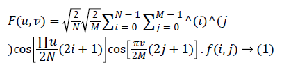
Discrete cosine transform
DCT is similar to the Fourier transform except that it uses only real numbers to specify the coefficients. DCT uses only fewer co-efficient to approximate lines, but DCT is not well suited for non stationary signals. The Discrete cosine transform and it is given by equation (1).
Discrete wavelet transform (DWT)
Discrete wavelet transform represent images in the time-scale domain. Wavelet transform consists of small wavelets with finite duration. By use of wavelet transform the image can be differentiated in to different frequency components with respect to time. Like Fourier transform in DWT different wavelets can be used as a basis function for representing other functions. Wavelet is represented as Ψ(x) which is the representation of the mother Wavelet and it is given in Eq. (2)
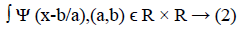
Where a=2-j and b=k.2-j, k and j are integers. The sampling of Lung image depends upon the value of a and b. DWT is very much suitable for point singularities, but they are not suitable for analyzing the images with geometric feature with line and surface singularity. So the feature extracted by DWT is not good for curves/edges of the lung region of the CXR. DWT Transform is applied to the segmented lung region and its coefficients are analyzed using statistical parameters.
Discrete curvelet transform
Curvelet transform is a multiscale pyramid transform that aims at a nonadaptive sparse representation of objects with edges. Curvelet transform has a highly directional sensitivity and anisotropy. Wavelet Transform has a less directional sensitivity when compared to curvelet. DWT is well suitable for analyzing point singularities, but not for edges/curves. There are four main steps in the curvelet transform [19].
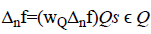
1. Sub band decomposition: In case of the sub band decomposition, image is filtered into a series of sub bands. Consider the segments lung region ‘f’ which is decomposed using the following relation
f=(P0Δ1f,Δ2f, Δ3f,……. . )
Where Δn is a window function, n=1,2,3…
2. Smooth partioning: The decomposed image is smoothly partitioned by using the window function it is given by
4. Ridgelet analysis: Now the renormalized function is analyzed using ridgelet analysis
Curvelet transform is applied to the segmented lung region and its coefficients are analysed using statistical parameters.
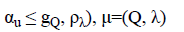
Discrete shearlet transform
DST is also one of the Multiresolution approaches. It is formed by scaling, shearing and translation. Shearlet transform gives the correct location and orientation of edges in the image. The discrete shearlet transform is obtained by sampling the continous shearlet transform [20], the discrete shearlet system and transform is given by equations (3) and (4). DST also provides a good anisotropic feature extraction.
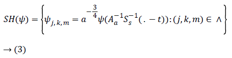
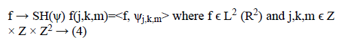
Discrete shearlet Transform is applied to the segmented lung portion and statistical parameters are calculated for shearlet coefficients.
Discrete bandelet transform
Bandelet transform is a self adaptive multiresolution geometric analysis which has the special properties of strict sampling and adaptability which are very important for image representation which is not present in the other MGA. It utilizes overall geometric information from the image which is more important in identifying every distinctive feature in the image. When compared to contourlet and curvelet transform, it provides stable and efficient performance [14]. Bandelet transform divides the image into sub blocks in multidirection and in multiscale, from which bandelet coefficients are generated. Bandelete transform is applied to the segmented lung region in our work and the statistical parameters are calculated from the bandelet coefficients.
Result and Discussion
In our work, the CXR image is processed in four steps to extract the feature from the lung region (Figure 2a). The first step in our method is image enhancement using CLAHE which is shown in Figure 2b for sample CXR image from JSRT database, then Watershed Segmentation is applied to segment the lung region from the CXR image which is shown in Figure 2c., and the features are extracted using five different transform such as DCT, DWT, DCRT, Shearlet and Bandelet. The features extracted from this transform are analysed using various statistical parameters for both Lung nodule (Normal) and Non-nodule (abnormal) images. Here we consider four statistical Parameters like Mean, Median, Energy and Entrophy. The comparison of this parameters for various transform both for normal and abnormal image is given in Table 1.
| DCT | DWT | DCRT | Shearlet | Bandelet | ||||||
|---|---|---|---|---|---|---|---|---|---|---|
| Normal | Abnormal | Normal | Abnormal | Normal | Abnormal | Normal | Abnormal | Normal | Abnormal | |
| Mean | 0.63 | 0.556 | 0.512 | 0.736 | 0.5875 | 0.864 | 0.166 | 0.438 | 0.302 | 0.636 |
| Median | 0.442 | 0.516 | 0.472 | 0.756 | 0.496 | 0.854 | 0.556 | 0.778 | 0.352 | 0.686 |
| Energy | 0.52 | 0.67 | 0.3384 | 0.6 | 0.272 | 0.728 | 0.256 | 0.502 | 0.43 | 0.572 |
| Entropy | 0.272 | 0.718 | 0.67 | 0.644 | 0.132 | 0.596 | 0.384 | 0.532 | 0.3682 | 0.652 |
Table 1: Comparison of Statistical Prameters for CXR images available in JSRT database.

Figure 2a: Input CXR image (JPCNN012.img).

Figure 2b: Enhanced image using CLAHE.

Figure 2c: Lung region segmented using watershed segmentation.
The analysis of statistical parameters for various images in the JSRT database is done graphicaly. The Graphical representation for mean of the Normal and abnormal image of the CXR is given in Figures 3a and 3b, median of the Normal and abnormal image of the CXR is given in Figures 3c and 3d, energy of the Normal and abnormal image of the CXR is given in Figures 3e and 3f, entrophy of the Normal and abnormal image of the CXR is given in Figures 3g and 3h.

Figure 3a: Mean for normal image.

Figure 3b: Mean for abnormal image.

Figure 3c: Median for normal image.

Figure 3d: Median for abnormal image.

Figure 3e: Energy for normal image.

Figure 3f: Energy for abnormal image.

Figure 3g: Entrophy for normal image.

Figure 3h: Entrophy for abnormal image.
In analyzing this various parameters and comparing the result produced by the different transform we suggest that both curvelet and Bandlet transform provide the features with good discrimination between the normal and abnormal images.
Conclusion and Future Work
CAD for automatic segmentation of Lung Region using Watershed Segmentation in CXR and feature extraction of the lung region using different transforms was developed. In our work we have extract the feature using DCT, DWT, DST, Curvelet and Bandelet Transform. The Feature extracted by this transforms are analyzed using the statistical parameters such as Mean, Median, Energy and Entropy. We have analyzed from the result of the Statistical Parameter for the various transform for Lung Nodule and Lung Non-Nodule image of the JSRT database, Curvelet and Bandelet Transform perform well. In our future work, we will use this transform for feature extraction of the Lung region for finding different abnormalities in the Lung Region.
References
- Chen S, Suzuki K. Separation of bones from chest radiographs by means of anatomically specific multiple massive-training anns combined with total variation minimization smoothing. IEEE Trans Med Imaging 2014; 33: 246-257.
- http://radiologymasterclass.co.uk/tutorials/chest/chest_quality/chest_xray_quality_projection
- Ma Z, Tavares JMRS, Jorge RMN. A review on the current segmentation algorithms for medical images. Proceedings of the First International Conference on Computer Imaging Theory and Applications. 2009.
- Shi Y, Qi F, Xue Z, Chen L, Ito K, Matsuo H, Shen D. Segmenting lung fields in serial chest radiographs using both population-based and patient-specific shape statistics. IEEE Trans Med Imaging 2008; 27: 481-494.
- Salam S. Level set segmentation method for automatic tuberculosis screening using chest radiographs. Int J Sci Res 2012; 3: 1633-1636.
- Dey EK, Muctadir HM. Chest X-ray analysis to detect mass tissue in lung. International Conference on Informatics, Electronics & Vision (ICIEV), 2014.
- Candemir S, Jaeger S, Palaniappan K, Antani SK, Thoma GR. Graph cut based automatic lung boundary detection in chest radiographs. 1st Annual IEEE Healthcare Innovation Conference of the IEEE EMBS Houston, Texas, USA, 2012.
- van Ginneken B, ter Haar Romeny BM, Viergever MA. Computer-aided diagnosis in chest radiography: a survey. IEEE Trans Med Imaging 2001; 20: 1228-1241.
- Candemir S, Jaeger S, Palaniappan K, Musco JP, Singh RK, Xue Z, Mcdonald CJ. Lung segmentation in chest radiographs using anatomical atlases with nonrigid registration. IEEE Trans Med Imaging 2014; 33: 577-590.
- Zhang H, Jiang X. An improved algorithm based on texture feature extraction for image retrieval. 12th International Conference on Fuzzy Systems and Knowledge Discovery (FSKD), 2015.
- Pun CM, Zhu HM. Textural image segmentation using discrete cosine transform. Proceeding CIT'09 Proceedings of the 3rd International Conference on Communications and Information Technology, 2009.
- Arivazhagan S, Kumar T, Ganesan L. Texture classification using curvelet transform. Int J Wavelets Multiresolut Inf Process 2007.
- Lim W. The discrete shearlet transform: A new directional transform and compactly supported shearlet frames. IEEE Trans Image Process 2010; 19: 1166-1180.
- Yang M. Feature vector extraction in hsv with bandlet transform. Proceedings of the 2nd International Conference on Intelligent Computing and Cognitive Informatics, 2015.
- Shiraishi J, Katsuragawa S, Ikezoe J, Matsumoto T, Kobayashi T, Komatsu K, Matsui M, Fujita H, Kodera Y, Doi K. Development of a digital image database for chest radiographs with and without a lung nodule: receiver operating characteristic analysis of radiologists' detection of pulmonary nodules. AJR Am J Roentgenol 2000; 174: 71-74.
- van Ginneken B, Stegmann MB, Loog M. Segmentation of anatomical structures in chest radiographs using supervised methods: a comparative study on a public database. Med Image Anal 2006; 10: 19-40.
- Sasi NM, Jayasree VK. Contrast limited adaptive histogram equalization for qualitative enhancement of myocardial perfusion images. Engineering 2013; 5: 326-331.
- Candemir S, Jaeger S, Palaniappan K, Antani S, Thoma G. Graph cut based automatic lung boundary detection in chest radiographs. 1st Annual IEEE Healthcare Innovation Conference of the IEEE EMBS, 2012.
- Candes EJ, Donoho DL. Curvelets, multiresolution representation, and scaling laws. Wavelet Appl Signal Image Process 4119: 408568.
- Kutyniok G, Labate D. Introduction to Shearlets. In: Applied and Numerical Harmonic Analysis. Springer, Berlin, 2012.