Research Article - Biomedical Research (2018) Volume 29, Issue 20
Breast cancer detection using image enhancement and segmentation algorithms
Yousif M.Y Abdallah1*, Sami Elgak1, Hosam Zain2, Mohammed Rafiq3, Elabbas A. Ebaid4 and Alaeldein A. Elnaema5
1Department of Radiological Science and Medical Imaging, College of Applied Medical Science, Majmaah University, Majmaah, 11952, Saudi Arabia
2Department of Obstetrics and Gynecology, College of Medicine, Majmaah University, Majmaah, 11952, Saudi Arabia
3Department of Public Health, College of Applied Medical Science, Majmaah University, Majmaah, 11952, Saudi Arabia
4Department of Radiology, College of Medicine, Majmaah University, Majmaah, 11952, Saudi Arabia
5Department of Surgery, College of Medicine, Majmaah University, Majmaah, 11952, Saudi Arabia
- *Corresponding Author:
- Yousif M.Y Abdallah
Department of Radiological Science and Medical Imaging
College of Applied Medical Science
Majmaah University, Saudi Arabia
Accepted date: November 21, 2018
DOI: 10.4066/biomedicalresearch.29-18-1106
Visit for more related articles at Biomedical ResearchAbstract
Enhancement of mammography images considers as powerful methods in categorization of breast normal tissues and pathologies. The digital image software gives chance to improve the mammographs and increasing their illustration value. The image processing methods in this paper were using contrast improvement, noise lessening, texture scrutiny and portioning algorithm. The mammography images kept in high quality to conserve the quality. Those methods aim to augment and hone the image intensity and eliminate noise from the images. The assortment factor of augmentation depends on the backdrop tissues and type of the breast lesions; hence, some lesions gave better improvement than the rest due to their density. The computation speed examined used correspondence and matching ratio. The results were 96.3 ± 8.5 (p>0.05). The results showed that the breast lesions could be improved by using the proposed image improvement and segmentation methods.
Keywords
Mammography, Image processing, Segmentation, Image enhancement, MatLab
Introduction
The breast lesion act as the one of the significant problems now a day, the first line diagnosable solution by detecting all lesions of the breast early to help and improving prognosis of cancer and other health problem related to breast [1-3]. More than one diagnostic modalities are used for breast cancer screening like computed tomography, resonance magnetic imaging and ultrasound. All of these modalities don’t enough to detect all lesions of the breast in spite of their characteristics such as high sensitivity of soft tissue detection. This shortening of detecting lead to support the final diagnosis decision by other further examinations like biopsy and unfortunately that may increase the anxiety of the patients [4-7]. The digital mammography is superior in detecting breast lesions than other diagnostic modalities. American cancer society recommended that MRI breast investigation should be mandatory for women with previous history of ovarian cancer or positive family history. Nowadays, a new method introduces to detect the beast lesions. This method is called digital mammography. Because its ability to link with image processing software [8-11]. Digital radiography acts as the useful modalities in lesions detection of the breast because its sensitivity and precision that may increase when used by well expert and trained radiologist. Many studies showed that 10-30% lesion of the breast were missing during routine screening [12].
The image processing plays an important role to decrease the cost of screening process by enhancing the result of digital mammography especially in case of small breast lesions. The first significant features of the breast cancer represent as mass with specific site, texture, border and shape, all these characteristics it easy to study under the image processing techniques by using the MATLAB program [14,15]. The idea of image processing by converting the result image into matrix as the binary image in addition to series of processes until reach to the appropriate result that helps to take a decision of diagnostic breast lesions [16,17]. There are many types of techniques used for mammography images detection such as edge-based transform, smoothing, noise removal and extraction techniques. Those methods help the physician in breast cancer diagnosis and monitor the treatment process [18,19].
The Research Method
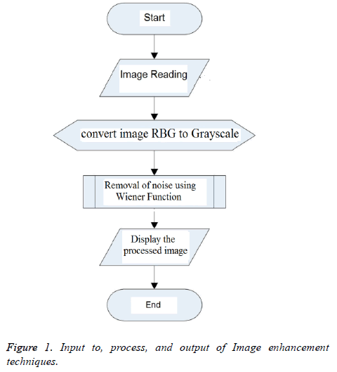
MatLab software used to implement the algorithm because its high-recital tool for image analysis and its assimilates subtraction. There are many algorithm, which used for contrast augmentation such as image adjustment, noise reduction using wiener transform. By execution those algorithm the intensity of image can be augmented or diminished which is useful in detection of some lesions. Furthermore, the color conversion can increase the lesion appearance and recognition of an object of curiosity. Mammographs are predisposed to diverse kind of noise. Those noise leads to blunders in the image gaining procedure that leads to pixel amounts that do not reflect the true intensities of real scene. The approach often procedure performed using Gaussian filtering as shown in Figure 1. Wiener estimates the local mean and variance around each pixel (Figure 1).

and

Where η is the N-by-M local region of respectively pixel in the mammograph.
ν2 represents the noise alteration.
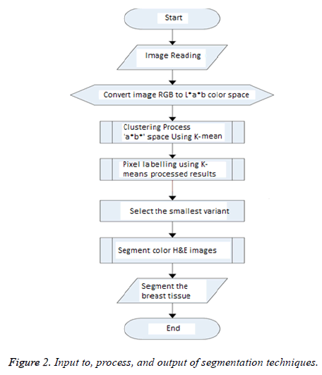
It is processing method used to categorize or subdivide an image into numerous parts by grouping the pixels according to their features. This paper accomplished to subdivision the mammography using texture scrutiny methods. The study comprised of different breast conditions. Initially, the breast boundaries itemized and segmented. Then segment the rest of breast soft tissue. Moreover, the mammograph exhibited within the full image as contextual image. Watershed transforms and colors study used to augment the mammographs images (Figure 2). The image recites into the program. In this study, forward algorithm used to change to RGB colour.
The forward algorism
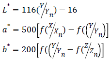
Where
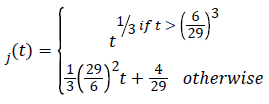
Where Xn, Yn, and Zn are the tristiulus values of the white point.
The breast images segmented automatically by using color analysis based technique (Figure 2).
Results and Discussion
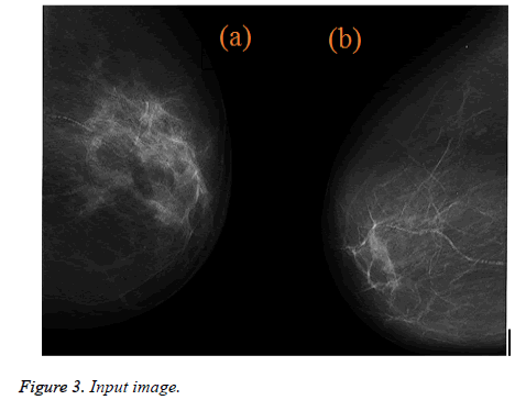
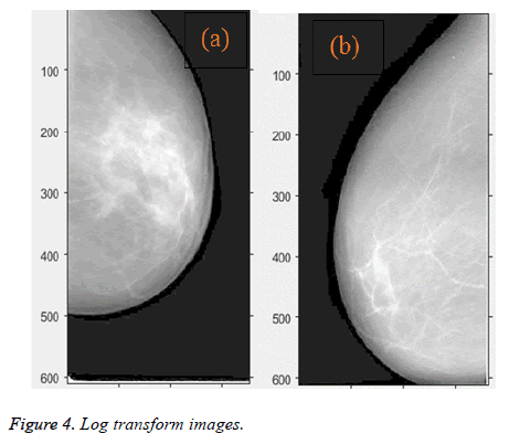
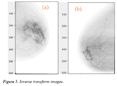
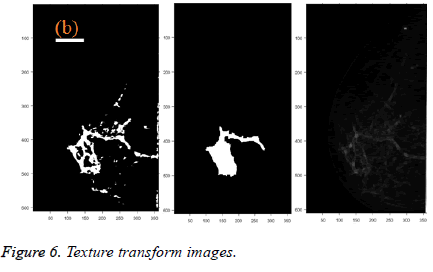
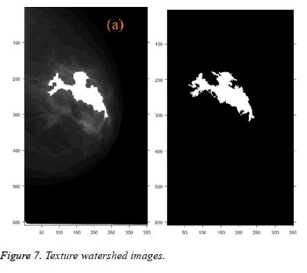
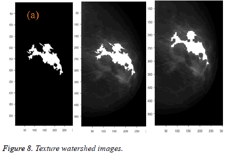
In pre-processing stage, the images read and saved as gray scale. Many filters were applied such as white and black filter, sharp image and log transformation. In this stage, the noise was removed followed by sharpening of the images and median filter application. The median and log transformation give best results comparing with the others. They work through mathematic equations to get rid the unwanted values of pixels and substituting them with wanted ones. They work in extemporized manner (non-linear) that increases the diagnostic and quality values of the images (Figures 3-8).
Abdallah [19], Mustra et al. [28] and Rahmati [29] suggested in their study that using the FCLAHE technique would increase the contrast and visibility of the images via noise and intensity heterogeneity reduction.
The results of this study showed that the suggested techniques have high matching and corresponding rate comparing with manual subjective analysis. Consequently, application of those techniques will reduce the time and improve the image in good manner.
It is noticeable that using of preprocessing techniques would improve that quality of images if associated with segmentation method. On the other hand, eliminating only noise is not adequate to attain 100% of precision.
Conclusion
Breast tissues segment using segmentation techniques. Texture analysis codes had developed especially for malignant tissues definition. The successfully algorithms used in this study gave high corresponding rate with less signal noise ratio (SNR) comparing with other scientist such as Qin [17], Abdallah [19] and Bushra et al. [21]. This technique is easy to apply unlike the other. Time saving, precision and reiterations are most important characteristics of this technique. In this study, we offered new techniques, which would help in detection of the tumor in mammography. Based on the results of the segmentation and improvement, we concluded the both methods would improve the images diagnostic value. The future works have to focus on and study detection of the all breast anomalies using other imaging modalities.
Acknowledgement
The authors are thankful to the Deanship of Scientific Research, at Majmaah University for funding this research.
References
- Liu J, Zhuang X, Wu L, An D, Xu J, Peters T, Gu L. Myocardium segmentation from de mri using multi-component gaussian mixture model and coupled level set. IEEE Trans Biomed Eng 2017; 1: 1-10.
- Abdallah Y. Application of analysis approach in noise estimation, using image processing program. Lambert Publishing Press GmbH & Co. KG 2011; 123-125.
- Abdallah Y, Yousef R. Augmentation of X-rays images using pixel intensity values adjustments. Int J Sci Res 2015; 4: 2425-2430.
- Abdallah Y. Increasing of edges recognition in cardiac scintography for ischemic patients. Lambert Publishing Press GmbH & Co. KG 2011; 123-125.
- Liu Y, Li C, Guo S, Song Y, Zhao Y. A novel level set method for segmentation of left and right ventricles from cardiac MR images. Conference Proceedings IEEE Engineering Medicine Biology Society 2014; 47: 19-22.
- Feng C, Zhang S, Zhao D, Li C. Simultaneous extraction of endocardial and epicardial contours of the left ventricle by distance regularized level sets. Med Phys 2016; 43: 2741.
- Ammar M, Mahmoudi S, Chikh MA, Abbou A. Endocardial border detection in cardiac magnetic resonance images using level set method. J Digit Imaging 2012; 25: 294-306.
- Li S, Yanping L, Kaitao Y, Shaozi L. ECG analysis using multiple instance learning for myocardial infarction detection. IEEE Transactions on Biomedical Engineering 2012.
- Wang L, Chitiboi T, Meine H, Günther M, Hahn HK, Principles and methods for automatic and semi-automatic tissue segmentation in MRI data. MAGMA 2016; 29: 95-110.
- Islam MK, Haque A, Tangim G, Ahammad T, Khondokar M. Study and analysis of ECG signal using MATLAB and LABVIEW as effective tools. Int J Comp Electric Eng 2012; 4.
- Lu X, Yang R, Xie Q, Ou S, Zha Y, Wang D. Nonrigid registration with corresponding points constraint for automatic segmentation of cardiac DSCT images. Biomed Eng Online 2017; 16: 39.
- Zhao H, Yan J. The wavelet decomposition and reconstruction based on the MATLAB. Proceedings of the Third International Symposium on Electronic Commerce and Security Workshops (ISECS) 2010; 10: 143-145.
- Qin X, Cong Z, Fei B. Automatic segmentation of right ventricular ultrasound images using sparse matrix transform and a level set. Phys Med Biol 2013; 58: 7609-7624.
- Qin X, Wang S, Shen M, Zhang X, Lerakis S, Wagner MB, Fei B. Register cardiac fiber orientations from 3D DTI volume to 2D ultrasound image of rat hearts. Proc SPIE Int Soc Opt Eng 2015; 9415-9419.
- Dormer J, Qin X, Shen M, Wang S, Zhang X, Jiang R, Wagner MB, Fei B, Determining cardiac fiber orientation using FSL and registered ultrasound/DTI volumes. Proc SPIE Int Soc Opt Eng 2016; 9790.
- Abdallah Y, Hassan A. Segmentation of brain in MRI images using watershed-based technique. Int J Sci Res 2015; 4: 683-688.
- Qin X, Wang S, Shen M, Lu G, Zhang X, Wagner MB, Fei B. Simulating cardiac ultrasound image based on MR diffusion tensor imaging. Med Phys 2015; 42: 5144-5156.
- Muhammad N, Bibi N, Jahangir A, Mahmood Z. Image denoising with norm weighted fusion estimators. Patt Anal Appl 2017; 1-10.
- Abdallah Y. Increasing of edges recognition in cardiac scintigraphy for ischemic patients. J Biomed Eng Med Imag 2016; 2.
- Muhammad N, Bibi N, Qasim I, Jahangir A, Mahmood Z. Digital watermarking using Hall property image decomposition method. Patt Anal Appl 2017; 1-16.
- Bushra M, Nazeer M, Muhammad S, Tanzila S, Amjad R. Extraction of breast border and removal of pectoral muscle in wavelet domain. Biomed Res 2017; 28: 1-3.
- Abdallah Y. Segmentation of heart tissues using gathering and colour analysis techniques. Biomed Res India 2017: 28: 1-4.
- Ma Y, Lu X, Dong M, Wang K. Automatic mass segmentation method in mammograms based on improved VFC snake model. Neur Networks Princip Appl 2014.
- Raba D, Oliver A, Mart J, Peracaula M, Espunya J. Breast segmentation with pectoral muscle suppression on digital mammograms. Patt Recogn Image Anal 2005; 471-478.
- Zhang Z, Lu J, Yip YJ. Automatic segmentation for breast skin-line. 10th IEEE International Conference on Computer and Information Technology 2010; 1604: 1599.
- Chen Z, Zwiggelaar R. A combined method for automatic identification of the breast boundary in mammograms. Biomed Eng Inform (BMEI) 2012 5th International Conference 2012; 125.
- Wei K, Guangzhi W, Hui D. Segmentation of the breast region in mammograms using watershed transformation. Eng Med Biol Soc 2005.
- Mustra M, Bozek J, Grgic M. Breast border extraction and pectoral muscle detection using wavelet decomposition. EUROCON 2009; 1426-1433.
- Rahmati P, Hamarneh G, Nussbaum D, Adler A. A new preprocessing filter for digital mammograms. Image Sig Proc 2010; 585-592.
