- Biomedical Research (2015) Volume 26, Issue 1
Severe hyperkalemia leads to bilateral calf pain and ECG abnormality in a patient with pacemaker.
Bingzhang Jie* , Ming Yang, Ling Han, Yan Zhao, Wenze HuDepartment of Cardiology, Fuxing Hospital, Capital Medical University, Beijing 100038, China.
- *Corresponding Author:
- Bingzhang Jie
Department of Cardiology
Fuxing Hospital Capital Medical University
Beijing 100038 China.
Accepted date: November 17 2014
Abstract
Hyperkalemia is an electrolyte abnormality that may cause cardiac pacemaker malfunction. Clinically, bilateral calf pain is extremely rare in hyperkalemia. Here we presented a case of a 78-year-old female who had bilateral calf pain with electrocardiographic changes in severe hyperkalemia.
Keywords
Hyperkalemia, bilateral calf pain, electrocardiographic changes, pacemaker.
Introduction
Hyperkalemia is a common clinical condition that could induce deadly cardiac arrhythmias, and is usually detected by electrocardiographic manifestations such as classic sinewave rhythm, and nonspecific repolarization abnormalities. However, sometimes it is difficult to diagnose hyperkalemia [1]. Hyperkalemia related bilateral calf pain is rarely reported. Here we presented a case of a 78-yearold female who had severe hyperkalemia with bilateral calf pain, and demonstrated electrocardiographic changes.
Case report
A 78-year-old female presented to the vascular surgery clinic because of progressive bilateral calf pain and cramping that began 3 days ago. She had no other complaints, except fatigue, nausea, upper abdominal pain and partial loss of hearing. She had no previous history of bilateral calf pain, with medical history of coronary artery disease, hypertension, and atrial fibrillation, which had been first diagnosed in 1992. Renal inadequacy diagnosed in 2009 (the highest creatinine value was 217 μmol/L). She received single ventricular pacemaker 10 years ago. Cardiac therapy consisted of spironolactone (20 mg daily), irbesartan (150 mg/d), and metoprolol. Her blood pressure was maintained at 110/70 mmHg.
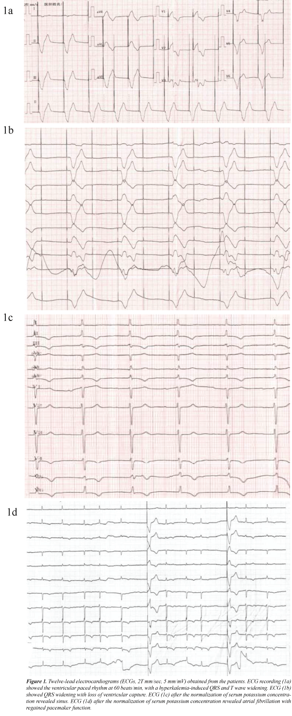
On admission, physical examination revealed that her limbs were pale and clammy, her blood pressure was 90/60 mmHg, and ECG showed regular pacing of pacemaker and widened QRS complex (Fig. 1a). A color doppler ultrasound of lower limbs arteries showed that bilateral popliteal artery had moderate stenosis, but the blood flowed smoothly. Since her symptoms persisted, she was asked to stay in the observation room. Six hours later, the patient developed excruciating pain and extreme weakness and sudden onset of hypotension and bradycardia. ECG showed that the pacemaker failed with further widened QRS complexes, the heart rate was 30 beats/min (Fig. 1b). Therefore, she was transferred to the emergency room. Physical examination revealed her inability to move limbs and her blood pressure dropped to 70/50 mmHg. Possibility of electrode dislocation verified. Preparation for temporary pacing was processed immediately. Rapid blood gas analysis showed that serum potassium was 9.9 mM/L, suggesting that hyperkalemia led to the failure of pacemaker capture. Isoproterenol, calcium gluconate, sodium bicarbonate, and insulin were administered to improve both the hemodynamics and the serum potassium, followed by emergency hemodialysis. After normalization of the serum potassium concentration, her cardiac rhythm became sinus rhythm and her heart rate was increased to 72 beats/min (Fig. 1c). The bilateral calf pain and quadriplegia resolved completely. She was discharged after one week when the serum potassium was 4.7 mmol/L. Repeated ECG recording showed regaining of pacemaker function (Fig. 1d).
Figure 1: Twelve-lead electrocardiograms (ECGs, 25 mm/sec, 5 mm/mV) obtained from the patients. ECG recording (1a) showed the ventricular paced rhythm at 60 beats/min, with a hyperkalemia-induced QRS and T wave widening. ECG (1b) showed QRS widening with loss of ventricular capture. ECG (1c) after the normalization of serum potassium concentration revealed sinus. ECG (1d) after the normalization of serum potassium concentration revealed atrial fibrillation with regained pacemaker function.
Discussion
In most cases, bilateral calf pain may involve a variety of etiologies, including musculoskeletal and vascular pathology such as deep venous thrombosis [2]. Hyperkalemia results in acute onset of quadriparesis or paralysis [3,4]. However, there are reports of leg pain induced by hyperkalemia, especially for the patients who underwent pacemaker implantation. Hyperkalemia is an electrolyte abnormality that may cause cardiac pacemaker malfunction. This case illustrated some typical ECG changes due to hyperkalemia during paced rhythm, including gradual widening of the paced QRS complex and eventual loss of ventricular capture that reverted with the correction of hyperkalemia [5]. As demonstrated in this case, initial characteristic electrocardiographic abnormalities in hyperkalemia during paced rhythm are widened QRS complexes, followed by the failure of pacemaker capture, which resulted in hypotension and bradycardia. Rapidly progressing hemodynamic instability on the basis of atherosclerotic stenosis may result in acute aggravating limb ischaemia, followed by bilateral calf pain and quadriparesis. Hyperkalemia often leads to the appearance of a typical sine-wave pattern and may ultimately result in cardiac arrest and respiratory failure without timely treatment [6,7]. Furthermore, hyperkalaemia should be considered in the differential diagnosis of the failure of pacemaker capture.
Hyperkalemia is a medical emergency that can lead to severe consequences and needs urgent attention. Recognition of the combination of bilateral calf pain and electrocardiographic abnormalities as showed in this case will help early diagnosis and treatment of hyperkalemia. Although hyperkalemia–related bilateral calf pain is extremely rare, clinicians should always be aware of this potentially life-threatening situation.
Conflicts of interest
None to declare.
References
- Parham WA, Mehdirad AA, Biermann KM, Fredman CS. Hyperkalemia revisited. Tex Heart Inst J. 2006; 33:40-7.
- Shiver SA, Blaivas M. Acute lower extremity pain in an adult patient secondary to bilateral popliteal cysts. J Emerg Med 2008; 34: 315-8
- Wilson NS, Hudson JQ, Cox Z, King T, Finch CK. Hyperkalemia-induced paralysis. Pharmacotherapy 2009; 29: 1270-2
- Huntgeburth M, Laudes M, Burst V, et al. Acute cardiac arrest secondary to severe hyperkalemia due to autoimmune polyendocrine syndrome type II. Clin Res Cardiol 2011; 100: 379-82
- Bahl A, Swamy A, Jeevan H, et al. Ecg changes of hyperkalemia during paced rhythm. Indian Heart J 2009; 61: 93-4
- Pluijmen MJ, Hersbach FM. Images in cardiovascular medicine. Sine-wave pattern arrhythmia and sudden paralysis that result from severe hyperkalemia. Circulation 2007; 116: e2-4
- Freeman SJ, Fale AD. Muscular paralysis and ventilatory failure caused by hyperkalaemia. Br J Anaesth 1993; 70: 226-7
