 44 7897 074717
Case Report - Ophthalmology Case Reports (2026) Volume 10, Issue 1
Ocular involvement in a patient with Rickettsia typhi: Case report.
Nagalakshmi Narayana Swamy*
Department of Ophthalmology, Naruvi Hospitals, Vellore, Tamil Nadu, India
*Corresponding Author:
- Nagalakshmi Narayana Swamy
Department of Ophthalmology, Naruvi Hospitals, Vellore, Tamil Nadu, India
E-mail: nag1994narayan@gmail.com
Received: 20-Dec-2024, Manuscript No. OER-24-155921; Editor assigned: 25-Dec-2024, OER-24-155921 (PQ); Reviewed: 08-Jan-2025, QC No. OER-24-155921; Revised: 06-
Feb-2026, Manuscript No. OER-24-155921 (R); Published: 13-Feb-2026, DOI: 10.35841/oer-10.1.281
Citation: Swamy NN. Ocular involvement in a patient with Rickettsia typhi: Case report. Ophthalmol Case Rep. 2026;10(1):281
Abstract
Background: A case of retinochoroiditis with vitritis with optic disc edema caused by Rickettsia typhi in a patient with previous normal eye.
Case presentation: This is case of 17-year-old female with no previous eye disease, who was referred to our ophthalmology. Patient had presented with history of fever for 5 days associated with headache. She presented with symptoms of floaters and diplopia since the onset of fever. Examination revealed 1+anterior vitreous cells, small retinal lesions and optic disc edema. Fluorescein angiography indicated mild discs hyperfluorescence, and the clinically visible round punctate lesions on optical coherence tomography showed inner retinal hyper-reflective lesion with a depth till outer plexiform layer possibly suggestive of a retinitis lesion. Laboratory tests were normal positive Rickettsia typhi serology tests, hypophosphatemia and hypovitaminosis D. Treatment included systemic doxycycline, azithromycin and prednisone, with improvement of visual acuity, ocular symptoms, optical coherence tomography abnormalities and resolution of inflammation. Oral prednisolone was discontinued, and after two months, additional improvement was seen clinically, with preserved retinal structures on optical coherence tomography.
Conclusion: This study is a case retinochoroiditis with vitritis with optic disc edema as a rare ocular presentation of Rickettsia typhi. There is need of awareness among clinicians about the ocular symptoms following systemic illness. Diagnosis relies on seroconversion, with fluorescein angiography and optical coherence tomography aiding in assessment.
Keywords
Rickettsia typhi, Retinochoroiditis, Vitritis, Optical coherence tomography.
Introduction
Murine typhus is caused by Rickettsiae typhi. It is transmitted by the rat flea, Xenopsylla cheopis. Symptoms of murine typhus are non-specific and can mimic those of other infectious diseases, thus it is often unrecognized and often under-reported [1,2].
The most common clinical features of murine typhus include triad of fever, headache, and rash. The clinical presentation is normally mild, but disease can be severe and even fatal. The severity of murine typhus is related to age, race, and delayed diagnosis. Furthermore, it is linked to inflammatory processes affecting components of the eye, with involvement of posterior segment such as retina, choroid and vitreous.
Here we report a rare presentation of retinochoroiditis and vitritis with optic disc edema secondary to the unusual infection of Rickettsia typhi.
Case Presentation
In October 2024, a 17 year old female was referred to our ophthalmology unit with no history of previous ocular disease with complaints of diplopia and floaters in the left eye for 2 days. She presented with a 7-day history of fever with headache and a 2-day history of floaters in the left eye. She had history of fever for 7 days 101.3° F of fever, headaches, and night sweats before the onset of ocular symptoms and a mild rash that covered her entire body and resolved spontaneously within one day [3-5].
There was no significant familial history of inflammatory or rheumatologic diseases. She denied recent travel/contact with animals.
On examination, her best corrected visual acuity in the right eye visual acuity was 20/20 and in the left eye was 20/25 with normal intraocular pressure of 10 mmHg and 12 mmHg respectively. Slit lamp examination showed 1+cells in the anterior vitreous in the left eye and fundus examination in both eyes revealed bilateral disc elevation and blurred optic disc margins (Figure 1). In the left eye, there was a well-defined yellowish focus consistent with retinitis adjacent to the fovea. Similar findings were found in the right macula. Periphery was normal.
Fluorescein angiography showed bilateral mild optic disc hyperfluorescence without vascular leakage (Figure 2). The visual fields test yielded normal results [6].
Optical coherence tomography
Right eye: Central foveal thickness was 321 μm, exhibiting a similar pattern but without outer band irregularities (Figure 3). Left eye: Central foveal thickness of 290 μm, with preserved contour and without cystic changes, and choroidal thickening noted (> 410 μm) Focal retinal hyperreflectivity was noted para-centrally, with irregularities in the photoreceptor layer. A hyperreflective foci was noted in the posterior vitreous also. Investigations found an elevated ESR (35 mmHr), negative for syphilis, toxoplasmosis serology and positive Rickettsia typhi serology. And was treated with doxycycline 100 mg twice daily for 4 weeks and prednisone 50 mg daily [7]. Two weeks following treatment the patient improved symptomatically. Visual acuity improved to 20/20 in the left eye too. Slit lamp examination revealed mild lower vitritis (0.5 +cells) in the left eye and fundus in the left eye showed, the macula appeared dry with two small, pinpoint, paracentral welldefined hypopigmented foci and normal peripheral retina. In the right eye, a dry macula with a small hypopigmented focus was located nasally to the fovea, and there was no inflammation on the periphery (Figure 4). She was advised to discontinue prednisone with further follow-up in our ophthalmology unit.
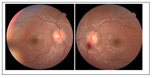
Figure 1. Showing optic disc edema with retinal hemorrhage with inflammatory cells in the vitreous. There was a small yellowish small focus consistent with retinitis adjacent to the fovea.
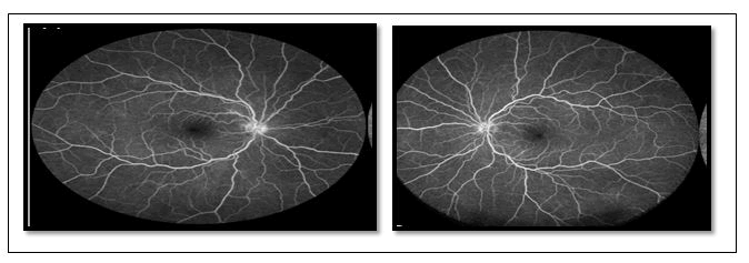
Figure 2. Bilateral mild disc hyperfluorescence without vascular leakage.
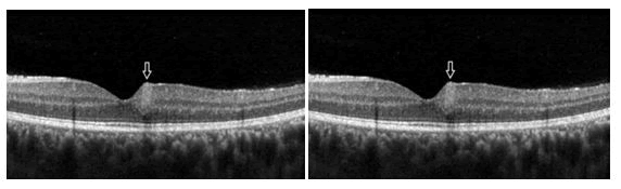
Figure 3. Hyperreflective retinal areas para-centrally, with irregularities in the photoreceptor layer. A small hyperreflective foci was noted in the posterior vitreous with choroidal thickening.
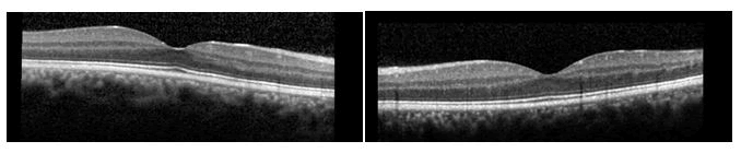
Figure 4. Prompt improvement was noted.
Discussion
Rickettsial disease have been prevalent worldwide and systemic rickettsial infection is now an emerging disease in India. Rickettsiae are obligate, intracellular, gram-negative bacilli residing in the arthropods such as lice, fleas, ticks and mites. Humans infection occurs after a bite by above stated insects and the bacilli then infect the vascular endothelium and reticuloendothelial cells. Rickettsial diseases are challenging due to non-specific systemic symptoms such as headache, fever, myalgia. Maculopapular skin rash and presence of eschar may aid in diagnosis of rickettsial disease. A positive history of recent forest visits, insect bite, zoonosis may aid the diagnosis of this infection [8]. Ocular involvement of rickettsia is known to present as conjunctivitis, vitritis, white retinal lesions and vascular leakage, post-infectious optic neuropathy and uveitis. Khairallah et al. showed retinitis in patients with rickettsia conorii infection. Here we report a case of vitritis, retinal involvement and choroidal involvement as supported by optical coherence tomography Khairallah M, et al. showed similar choroidal involvement in their study. Majority of the patients with ocular involvement remain asymptomatic although many may present with decreased vision, redness and floaters. In the study done by Khairallah M et al. in 2004, 20% had unilateral and 80% bilateral ocular involvement; out of which 48.3% had retinal vasculitis, 30% had retinitis, 1.3% had cystoid macular edema, 1.3% had optic disc edema. In the study done by Khairallah M et al. in 2009, 22% had unilateral and 78% had bilateral ocular involvement, out of which 42.5%had retinitis, 21.3% had vascular sheathing, 2.1% had macular edema and 4.3% had optic disc edema. The gold standard method of diagnosis is defined as a fourfold increase in serologic IgM and IgG antibodies to Rickettsia typhi during the convalescent phase, detected using Immunofluorescence Assay (IFA). Polymerase Chain Reaction (PCR) assays of anterior chamber fluid may be considered for
References
- Raoult D, Roux V. Rickettsioses as paradigms of new or emerging infectious diseases. Clin Microbiol Rev. 1997;10(4):694–719.
[Crossref] [Google Scholar] [PubMed]
- Walter G, Botelho-Nevers E, Socolovschi C, et al. Murine typhus in returned travelers: a report of thirty-two cases. Am J Trop Med Hyg. 2012;86(6):1049–53.
[Crossref] [Google Scholar] [PubMed]
- Masalha R, Merkin-Zaborsky H, Matar M, et al. Murine typhus presenting as subacute meningoencephalitis. J Neurol. 1998;245(10):665–8.
[Crossref] [Google Scholar] [PubMed]
- Abroug N, Khochtali S, Kahloun R, et al. Ocular manifestations of rickettsial disease. J Infect Dis Ther. 2014;2:140.
- Kawali A, Mahendradas P, Srinivasan P, et al. Rickettsial retinitis-an Indian perspective. J Ophthalmic Inflamm Infect. 2015;5:37.
[Crossref] [Google Scholar] [PubMed]
- Sankar S, Kulkarni S, Manjoo CS, et al. Rare ophthalmic manifestations of rickettsial infections. Int J Adv Res. 2017;5:1027–31.
- Rathi N, Rathi A. Rickettsial infections: Indian perspective. Indian Pediatr. 2010;47:157-164.
[Crossref] [Google Scholar] [PubMed]
- Zhang J, Pau D, Lee AG. Postinfectious optic neuropathy in endemic typhus. J Neuroophthalmol. 2011;31(4):342–3.
[Crossref] [Google Scholar] [PubMed]
- Khairallah M, Ben Yahia S, Toumi A, et al. Ocular manifestations associated with murine typhus. Br J Ophthalmol. 2009;93(7):938–42.
[Crossref] [Google Scholar] [PubMed]