Case Report - Otolaryngology Online Journal (2017) Volume 7, Issue 2
Nasal neuroglial heterotropia in a 14 year old boy
- *Corresponding Author:
- Ruqaiya Shahid, Assistant Professor of Pathology, Dow International Medical College, Dow University of Health Sciences, Ojha campus, C-88, block A, North Nazimabad, Karachi, Pakistan, Tel: 03323047919, E-mail: ruqaiyashahid@yahoo.com
Received date: March 09, 2017; Accepted date: March 30, 2017; Published date: April 07, 2017
Abstract
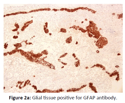
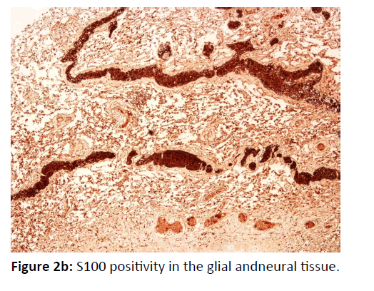
Nasal neuroglial heterotopia is a rare, congenital lesion, composed of mature brain and neural tissue that has isolated from the cranial cavity or spinal canal. It typically presents at birth or in early childhood. We describe a case of nasal neuroglial hetrotropia in a 14 year old boy who had a history of right nasal obstruction and discharge since birth. Anterior rhinoscopy showed an oval, elastic, smooth and uncompressible mass at the upper third of the nasal septum. He underwent surgery with a preoperative diagnosis of nasal cyst. Histologically, the lesion showed mature glial and neural tissue in a loose fibrous stroma. The glial tissue was strongly positive for immunohistochemical (IHC) antibody Glial Fibrillary Acidic Protein (GFAP). The neural tissue was positive for S100 antibody. We are reporting the case because of its rarity, particularly in teen age, and to emphasize the importance of immunohistochemistry in reaching a correct diagnosis.
Keywords:
Glial heterotropia; Nasal glioma; Nasal obstruction
Introduction
Nasal neuroglial heterotopia also called nasal glioma is a rare entity and was first described by Reid in 1852 [1]. The reported incidence of congenital nasal masses is 1 in every 20,000 to 40,000 births, and nasal neuroglial heterotropia is one of the common ones [2]. Neuroglial heterotropia are of two types: intranasal and extra nasal. Sixty percent are extranasal, 30% are intranasal, and 10% are both [3]. Nasal glioma is a misnomer as this term implies a neoplastic lesion of glial tissue, however; these lesions are heterotropic masses of neurogenic origin, which have lost their intracranial connection, and present as an obvious external or intranasal mass at birth [4]. Extranasal lesions at the dorsum of the nose usually cause a cosmetic deformity and intranasal lesions cause upper airway obstruction, respiratory distress and nasal discharge [5]. The treatment of choice is surgical excision and few recurrences are reported [6].
Case Report:
A 14 year old boy presented with a history of right sided nasal obstruction and nasal discharge since birth. He had had several ear infections and had been treated with multiple antibiotics. The family history and sibling history was unremarkable.
Physical examination showed an alert young boy, with mucoid nasal discharge and teary eyes. Anterior rhinoscopy showed an oval, elastic, smooth, uncompressible mass at the upper third of the right nasal septum, and the nasal septum was deviated to the left. The mass did not bleed to touch. Computed tomography (CT scan) confirmed a mass in the nasal cavity with a solid and cystic component and with pressure effects on the right inferior and middle turbinates. There was no infiltration into the surrounding planes and no intracranial extension. Considering preliminary differential diagnoses of a retention cyst and nasal polyp; the mass was excised.
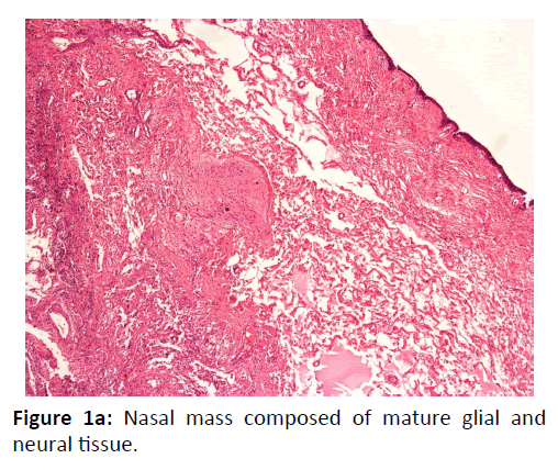
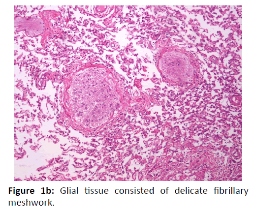
On gross examination, the mass was irregular, grayish-white and measured 4.5 × 2 × 1.5 cm. On cross section, the mass was elastic and smooth with a cyst measuring 1.5 × 1 cm. On microscopic examination, the nasal mass was composed of mature glial and neural tissue in fibro vascular and edematous stroma, covered by respiratory mucosa (Figure 1a). Glial tissue consisted of delicate fibrillary meshwork of astrocytes arranged in islands, some calcifications were present. Nerve fibers and neuritis were present in abundance (Figure 1a and 1b). Cystic degeneration was present. The glial tissue was positive for GFAP antibody (Figure 2a). S100 positivity was seen in the glial as well as neural tissue (Figure2b).
Patient is well, at three years of follow-up after surgery, with no recurrence.
Discussion
Nasal neuroglial heterotopia is a rare condition, characterized by the presence of heterotopic neuro glial tissue outside the nervous system [2]. It is thought to arise from displacement of neuroectodermal cells at an early stage of embrogenesis that may subsequently differentiate into a variety of cells of neuroglial tissue, or from protrusion of the neuro-glial tissue from the developing cerebral tissue that becomes isolated from the brain in later development [4,5]. Male-to-female ratio is 3:2. No familial predisposition has been described [6]. These lesions are mostly midline and the most frequently affected site is the nasal area, followed by the tongue, palate-pharyngeal complex, orbit, scalp, and ear[7]. As these are congenital lesions, the usual age of presentation of the nasal glial heterotropia is infancy or early childhood, but in this case the boy was diagnosed at 14 years. Case reports in teen age are rare in literature [8].
Nasal neuroglial heterotropia may have a connection with the intracranial glial tissue in 10-15% of cases, through a bone defect [8]. They are differentiated from an encephalocele which always has an intracranial connection and transilluminates on Valsalva test [8] .
Computed tomography and magnetic resonance imaging are complementary in determining the location, extent, relationship to the skull base, and for diagnosis and treatment planning [9]. Complete surgical excision is recommended and is curative [6]. Biopsy or fine needle aspiration of childhood nasal masses is contraindicated because of the increased risk of meningitis from ascending infection, in case of lesions with an intracranial connection [4].
Other differential diagnoses that should be considered are dermoid cyst, inflammatory nasal polyp, teratoma, nasal angiofibroma, meningioma, lymphangioma, rhabdomyosarcoma and hemangioma [10]. Histopathology is diagnostic and immunohistochemical staining is supportive in correctly identifying the neurogliual tissue in difficult cases [10].
Conclusion:
Nasal neuroglial heterotropia is not a true neoplasm and presents mostly in infants as recurrent nasal obstruction and upper respiratory infection. However, it may rarely present in teenage and must be considered in the differential diagnosis of airway obstruction.
References
- Al-Ammar AY, Al Noumas HS, Alqahtani M (2006) A midline nasopharyngeal heterotopic neuroglial tissue. J LaryngolOtol120:E25.
- Verney Y, Zanolla G, Teixeira R, Oliveira LC (2001) Midline nasal mass in infancy: A nasal glioma case report. Eur J PediatrSurg 11:324-327.
- Uzunlar KA, Osma ST, Yilmaz F, Topcus U (2001) Nasal glioma: Report of two cases. Turk J Med Sci 31:87-90.
- Penner CR, Thompson L (2003) Nasal glial heterotopia: Aclinicopathologic and immunophenotypicanalysis of 10 cases with a review of the literature. Ann DiagnPathol 7:354-359.
- Kamath S, Subbarao KC (2013)Nasopharyngeal glial heterotopia: A rare cause of airway obstruction in an infant. Indian J PatholMicrobiol 56:62-63.
- Talwar OP, Pradhan S, Swami R, KCSR (2007) Nasal glioma: A case report.Kathmandu Univ Med J (KUMJ) 5:11411-5.
- Sun LS, Sun ZP, Ma XC, Li TJ (2008) Glial choristoma in the oral and maxillofacial region; Aclinicopathologic study of 6 cases. Arch Pathol Lab Med 132:984-988.
- Daffon DVF, Calderon AF, Victoria FA (2013) Nasal Glial Heterotopia: Unsuspected brain tissue in the nasopharynx. Philipp J Otolaryngol Head Neck Surg 28:18-21.
- Shah J, Patkar D, Patankar T, Krishnan A, Prasad S, et al. (1999) Pedunculated nasal glioma: MRI features and review of the literature. J Postgrad Med 45:15-17.
- Gnagi SH, Schraff SA (2013) Nasal obstruction in newborns. Pad Clin N Am 60:903-922.


