Case Report - Current Pediatric Research (2023) Volume 27, Issue 7
Myocutaneous cysticercosis in a 2-year-old girl: A rare case report.
Mohammad Tahazzul*, Raj Wardhan, Motamarri Naga Sai Manikya Deepu, D Piyush Jain
1Department of Pediatrics, TS Misra Medical College and Hospital, Lucknow, UP, India
- Corresponding Author:
- Mohammad Tahazzul
Department of Pediatrics, TS Misra Medical College and Hospital, Lucknow, UP, India
E-mail: tahazzul@gmail.com
Received: 28 June, 2023, Manuscript No. AAJCP-23-105028; Editor assigned: 30 June, 2023, Pre QC No. AAJCP-23-105028(PQ); Reviewed: 14 July, 2023, QC No. AAJCP-23-105028; Revised: 21 July, 2023, Manuscript No. AAJCP-23-105028(R); Published: 31 July, 2023, DOI:10.35841/0971-9032.27.07.1956-1958.
Abstract
Cysticercosis is caused by the larval stage of the tapeworm (Taenia solium). Humans can be infected either by consumption of improperly cooked pork or by consumption of improperly washed food contaminated with the eggs of the tapeworm. Although the larva most commonly affects the central nervous system and orbits but it can involve various other organs like muscles, lungs, liver, heart and subcutaneous tissues. It can present with variable clinical manifestations according to the organ involved. Here we report a case of a two-year-old girl with myocutaneous cysticercosis involving the right lumbar region. The patient was managed with a course of steroids initially followed by oral albendazole.
Keywords
Cysticercosis, Myocutaneous cysticercosis, Taenia solium, Abdominal swelling, Ultrasound.
Introduction
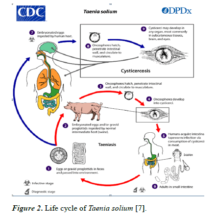
Cysticercosis is a common infection caused by the larval stage of tapeworm (Taenia solium). The parasite can cause two different types of infection in children [1]. Children can be infected with the tapeworm form by the consumption of improperly cooked pork which contains the larval cysts. After consumption, the cysts are converted into the tapeworm form in the intestines. Children can also acquire the infection by the eggs shed by the tapeworm carriers.
After the ingestion of eggs, the larvae are released from the eggs and invade the intestines and enter bloodstream. From the bloodstream they migrate to the muscles and other organs where tissue cysts are formed. Infection with the cystic form is known as cysticercosis whereas involvement of the central nervous system is called neurocysticercosis. The tapeworm form of the infection develops only after the ingestion of undercooked pork. While ingestion of pork is not essential to develop cysticercosis but individuals who harbour the adult form may infect themselves with the eggs by the fecal-oral route.
The clinical syndromes caused by the larval stage of tapeworm include neurocysticercosis and extra neural cysticercosis [2]. Cysticercosis is endemic in northern India, especially in the states of Bihar, Uttar Pradesh, and Punjab. A recent study on a rural pig farming community of Mohanlalganj block, Lucknow district of Uttar Pradesh, has reported the incidence of taeniasis at 18.6% [3].
Cysticercosis is widely prevalent among swine population in India. The Mohanlalganj area in Lucknow, where pig rearing is prevalent, has reported a high incidence of cysticercosis (26%) in swine, with more than 40% of them having NCC [4,5].
Cysticercosis is one of the most common causes of adult-onset seizures accounting for one third of the total cases. Paediatric cases of extra neural cysticercosis have hardly been reported. Here we report a case of a 2 year-old girl with a solitary swelling of the right lumbar region. There was no neurological or ocular involvement.
Case Presentation
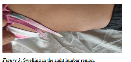
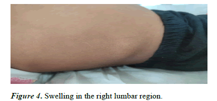
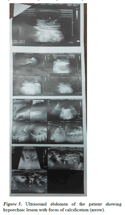
A 2 year old girl, on a vegetarian diet presented to the outpatient department with a painless solitary swelling in the right lumbar region measuring 2 cm × 1 cm. The swelling gradually increased in size over a period of one month. On examination the swelling was mobile, non-tender, well defined, oval shaped, non-reducible, firm in consistency and nonfluctuating. There was no history of fever or discharge from the swelling. There was no local rise of temperature, erythema or pain. It was a solitary swelling with no similar swellings in other parts of the body. The systemic examination was unremarkable. There was no history of trauma. The ultrasound of abdomen showed a well-defined hypoechoic lesion with focus of calcification measuring approximately 16 mm × 7 mm in size. The calcification was seen in the superficial plain of right lumbar region abutting the underlying muscle (external oblique).
With the clinical examination and the ultrasonography findings, a diagnosis of myocutaneous cysticercosis was made. The stool examination for ova and parasites was unremarkable. Chest radiograph and erect abdomen radiograph were unremarkable. FNAC of the swelling was done. On aspiration a white material and blood were collected. Smears showed sheets of neutrophils admixed with histiocytes and lymphocytes. No granuloma or malignancy was seen in the smears observed. A biopsy was suggested but the parents of the girl did not consent for the procedure. NCCT of the brain and ophthalmology evaluation were done and were found to be insignificant. As it was a case of a solitary non infected myocutaneous cysticercosis, conservative management was done with oral prednisolone for initial 3 days followed by a course of albendazole for 28 days (Figures 1-5) [6].
Figure 2: Life cycle of Taenia solium [7].
Results and Discussion
Cysticercosis is a parasitic infection affecting tissues caused by larval cysts of the tapeworm Taenia solium. These larval cysts can infect the brain, muscle, or other tissue, and are a major cause of adult-onset seizures (30%) in most low-income countries. A person gets cysticercosis by swallowing eggs found in the feces of a person who has an intestinal tapeworm [8]. Although vegetarians can also get the infection by ingestion of eggs from improperly washed vegetables. Infection can also be causes by the intake of undercooked pork meat. The tapeworm form of the infection develops only after the intake on undercooked pork.
Human cysticercosis can have devastating effects on human health. The larvae (cysticerci) may develop in the muscles, skin, eyes and the central nervous system [9]. Cysts are usually formed in the central nervous system and the eyes accounting for almost 86% of the cases. Other body parts that can be involved include the heart, lungs, peritoneum, breast, and muscles. Myocutaneous and subcutaneous forms of the disease are relatively rare specially in the paediatric population.
Conclusion
Although subcutaneous and myocutaneous forms of cysticercosis are very rare but it should be kept in mind as a differential diagnosis for a soft to firm abdominal swelling, especially in areas where cysticercosis is highly endemic. An ultrasound examination should be done as an initial diagnostic tool. FNAC/FNAB should be done for the confirmation of the diagnosis. Stool examination for ova and cysts should be done. Ophthalmology evaluation should be done to check for orbital spread. A CT/MRI of the brain should also be done for central nervous system involvement as CNS and ocular involvement are relatively more common. As cysticercosis is spread via undercooked and improperly washed food, proper hygienic practices should be encouraged and awareness regarding the disease should be generated in the public. Mass chemotherapy for tapeworm carriers, mass treatment of pigs, and improved personal hygiene have decreased or eliminated transmission in some areas.
References
- Clinton White A, Philip Fischer R. Cysticercosis. Nelson textbook of pediatrics. (1st edn) Elsevier 2020; 1895-1897.
- Clinton White Jr A, Christina Coyle M, Rajshekhar V, et al. Diagnosis and treatment of neurocysticercosis: 2017 clinical practice guidelines by the Infectious Diseases Society of America (IDSA) and the American Society of Tropical Medicine and Hygiene (ASTMH). Clin Infect Dis 2018; 66(8): e49.
- Prasad KN, Prasad A, Gupta RK, et al. Prevalence and associated risk factors of Taenia soliumtaeniasis in a rural pig farming community of north India. Trans R Soc Trop Med Hyg 2007; 101: 1241–1247.
- Prasad KN, Chawla S, Jain D, et al. Human and porcine Taenia solium infection in rural north India. Trans R Soc Trop Med Hyg 2002; 96(5): 515–6.
- Ahmad R, Khan T, Ahmad B, et al. Neurocysticercosis: A review on status in India, management, and current therapeutic interventions. Parasitol Res 2017; 116: 21–33.
- Prasad KN, Prasad A, Verma A, et al. Human cysticercosis and Indian scenario: A review. J Biosci 2008; 33:571–580.
- Centers for disease control and prevention. Cysticercosis. DPDx-Laboratory identification of parasites of public health concern.
- World Health Organization. Taeniasis/cysticercosis. 2022.
- Bhat V, Nagarjuna M, Belaval V, et al. Cysticercosis of the masseter: MRI and sonographic correlation. Dentomaxillofac Radiol 2015; 44(5): 20140372.
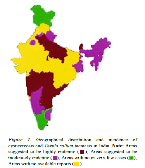
 ); Areas suggested to be moderately endemic (
 ); Areas with no or very few cases (
 ); Areas with no available reports (
 ).
).