Research Article - Ophthalmology Case Reports (2017) Volume 1, Issue 1
Modification of block matches three dimension algorithms for de-noising spatial domain optical coherence tomography images.
Kamalakshi N1*, Shanmukh Swamy N2
1Department of Computer Science and Engineering, University of Mysore, India
2Department of Electronics & Communications, Mysore, India
- *Corresponding Author:
- Kamalakshi N
Department of Computer Science and Engineering
University of Mysore, India
Tel: +91-821-2419334
E-mail: kamalnags@gmail.com
Accepted date: June 20, 2017
DOI: 10.35841/ophthalmology.1.1.47-50
Visit for more related articles at Ophthalmology Case ReportsAbstract
The analysis in a therapeutic picture assumes an imperative part highlighting on operations as picture rebuilding, include extraction and protest acknowledgment. These operations may end up plainly chaotic if the pictures are debased with clamors. So formulating a proficient calculation for clamor evacuation is a vital and testing research range in nowadays even with plenteous existing de-noising calculation. Creating Image de-noising calculations is a bulky assignment since vital subtle elements in a therapeutic picture inserting symptomatic data ought not be destroyed while expelling the commotion. This paper proposes an altered de-noising approach for retinal Optical Tomography Images by fusing un-annihilated Wavelet Transform to the current piece coordinating three measurement calculation, which that its uses in The outcomes demonstrates that the de-noised picture utilizing adjusted BM3D(Block Matching three measurement) have an enhanced PSNR and visual quality when contrasted with the condition of craftsmanship BM3D calculation which in turns will help the specialists to finding the malady better.
Keywords
De-noise, speckle noise, optical coherence tomography.
Introduction
Optical intelligibility tomography (OCT) is a rising imaging methodology that has been broadly utilized as a part of the field of biomedical imaging. In the current past, it has discovered uses as a demonstrative apparatus in dermatology, cardiology, and ophthalmology. In this paper we concentrate on its applications in the field of ophthalmology and retinal imaging. OCT can nonobtrusively create cross-sectional volumetric pictures of the tissues which can be utilized for investigation of tissue structure and properties. Because of the hidden material science, OCT pictures experience the ill effects of a granular example, called dot commotion, which limits the procedure of translation. This requires particular clamor decrease strategies to dispense with the commotion while protecting picture points of interest.
Optical intelligibility tomography (OCT) is a capable imaging framework for procuring 3D volumetric pictures of tissues non-intrusively. In basic terms, OCT can be considered as echography with light [1,2]. Dissimilar to echography which is finished by sound waves, OCT imaging is not time-of-flight based but instead produces the picture in view of the impedance designs demonstrates a run of the mill retinal OCT picture with false shading. All through the previous two decades, new advancements in the OCT imaging framework have enhanced the procurement time and furthermore the nature of the obtained pictures. These days taking (μm-level) volume pictures of the tissues is extremely normal particularly in ophthalmology and retinal imaging. Because of the volume of information produced in a clinical setting, there is a requirement for hearty and mechanized investigation methods to completely use the abilities of OCT imaging [3].
In the previous decade thorough and comprehensive research has been done both in the fields of bio restorative imaging and remote detecting for smothering spot commotion. Copious methodologies have been contrived to upgrade the picture quality corrupted by dot commotion [4,5] . A few dot lessening methods are depicted by [6,7]. A wavelet based delicate thresholding procedure has been beforehand connected to OCT pictures adulterated by dot commotion [8]. It registers the undecimated wavelet change and applies delicate thresholding to the level, vertical and asks few sub groups. The edge is acquired utilizing the measurements of the wavelet coefficients. The wavelet based procedure portrayed in [8] does not lessen the picture sharpness altogether but rather the execution time for the calculation is around 7 min utilizing Matlab usage. Altered Lee and Kuan versatile channels have been connected to SAR spot lessening [9].
Anisotropic dispersion is one of eminent that has been before connected for spot commotion expulsion in OCT pictures. For instance, in references [6,7] the angle of the picture is utilized for the computation of the dissemination coefficient with no thought to the genuine clamor present. Bo Chong and Yong- Kai Zhu proposed a novel dot commotion decrease calculation in OCT. The calculation depends on piece coordinating 3D channel altered by morlet wavelet decomposition. Original OCT picture information changed by logarithmic pressure is deteriorated into 10 segments by morlet wavelet for three levels. Every part is proposed by a suited BM3D channel and the yield picture is recreated by wavelet turn around change [8]. Mashaly et al. exhibited a versatile numerical morphological channel is proposed to lessen the dot commotion in SAR pictures [9]. Optical Coherence Tomography Noise Reduction Using Anisotropic Local Bivariate Gaussian Mixture Prior in 3D Complex Wavelet Domain Speckle decrease in optical rationality tomography pictures by utilization of a spatially versatile wavelet filter [10]. Retinal OCT Image Enhancement through Wavelet De-noising A de-noising calculation utilizing twofold thickness double tree complex wavelet change is connected to 2-outline OCT pictures of the retina to give an equivalent picture quality in less procurement time contrasted with usually utilized numerous edge averaging technique. BM3D is a current de-noising technique in view of the way that a picture has a locally inadequate portrayal in change area. This scarcity is upgraded by gathering comparative 2D picture patches into 3D groups [11-14].
Materials and Methods
The spot commotion is flag subordinate in its property. Subsequently it is exceptionally repetitive to dispose of without losing vital picture data, similar to edges or surface. In principle, fix based strategies can create an ideal de-noising result for Gaussian clamor yet they are likely not the most reasonable techniques for lessening the spot commotion show in OCT imaging. In our proposed technique, we examined receiving a middle sifting venture with a specific end goal to make the clamor show in OCT pictures more like a Gaussian dispersion, and lessening its flag reliance [15-17].
The fundamental strides of proposed strategy are outfitted beneath:
1. Take an info boisterous OCT picture
2. Apply middle channel to it as a pre-processor
3. Next apply square coordinating to discover the places of the pieces in z that are like the as of now prepared one. Shape a 3D exhibit (gathering) by stacking the pieces situated at the got areas.
4. The fundamental idea of UDWT is that it applies the change at each purpose of the picture and spares the itemized coefficients and utilizations the guess coefficients for the following level. The measure of the coefficients exhibit does not lessen from level to level.
5. Apply the UDWT to the first picture to deliver wavelet coefficients for separating the conceivable coefficients. The first image (level 0) into one low recurrence band (low-high, highlow and high –high groups point by point coefficients) for every determination level with an indistinguishable size from the first picture. The decay are generally led up to determination level 4
6. This disintegration is additionally iterated up to level 4. In the wake of figuring UDWT picture, a thres holding of the definite coefficients at all levels prepared for hard edge and picture is reproduced by utilizing converse discrete wavelet change. On isolating the coefficients, the destitute low recurrence coefficients are handled by backwards un-destroyed wavelet change.
7. The yield picture of past stride is part into different pieces of standard size, which are contrasted with each other with ascertain euclidean separations.
8. The piece with euclidean separation lesser than the edge esteem is bolstered to un-devastated wavelet change and coefficients are removed.
9. The low recurrence coefficients removed are then .The recreated picture acquired in the last stride is sent to unannihilated wavelet change for by and by figuring the conceivable coefficients.
10. On low recurrence coefficients a Wiener channel is connected for handling and the opposite discrete wavelet change is connected on the coefficients for remaking the picture.
Noise model
The speckle noise present in an OCT images are multiplicative noise which is nothing but an unwanted random signal which gets multiplied with some significant signal in the image capturing, transmission or processing. Mathematically speckle noise can be represented as follows:
Where Dm, n is the noisy pixel, Sm, n is the noise free pixel, Vm, nis the multiplicative noise m, n are spatial locations.
Identifying a noisy pixel (based on current pixel & its neighborhood values)
Use a sliding window to identify the noisy pixel and restore it
Apply a median filter to reduce the noisy pixels from image before applying the actual de-noising method
Design
Accurate current pixels as Px, Compare each pixel with its neighboring pixels
If Px is corrupted
Px value is maximum or minimum of all other pixel values in the corresponding neighbor
Sort the pixel value except Px of the filter window w_(x,) ^ n in ascending order
The remaining steps are same as block match three dimension algorithm [5].
Un-decimated wavelet Transform
The Un-decimated Discrete Wavelet Transform has been independently developed by numerous researchers separately defined by a number of names including “algorithm a trouse, the Shift Invariant DWT (SIDWT), and “Discrete Wavelet Frames (DWFs) [18-20].
The un-decimated wavelet filter at scale l+1 is defined recursively as g (l+1) [k] =g (l) [k] ↑ 2=g (l) k 2, if k even 0, if k odd (1) .The down sampling at each stage of the DWT is removed to give the UDWT. The shift variation of the DWT is caused by this subsampling and its removal within the UDWT provides perfect shift invariance. Furthermore, each sub-band is now the same size as the original signal leading to a considerably over complete representation. The near shift invariance and improved directional selectivity have facilitated excellent results in denoising, fusion and other image processing applications.
Aj+1[n]=aj [n] *hl [n]
Aj+1[n]=aj [n] *hl [n] - [4]
Data Collection
A set of 10 clinical SD-OCT images were collected from 10 patients of Vijaya Nethralaya Bangalore acquired in patients diagnosed with acute macular degeneration of the retina to qualitatively assess the demising performance of our proposed method.
Each SD-OCT image was acquired and the other dataset were obtained from duke university database [10]
The rating for each images were done by assigning grades from 1 to 3, with lower grades indicating higher demising performance. We also computed the method noise (the image difference between the original image and the denied image for eacalsoh de-noising method as to evaluate the impact of the method on the preservation of the image structure information. An optimal demising method should produce a method noise containing as little structure information as possible, such as shape edges.
Experimental Studies to evaluate methods: The experiments were conducted on a machine of 2.83-GHz Pentium 4 PC with 3.37 GB memory. The implementation of each de-noising method was in Matlab and run on the same machine.
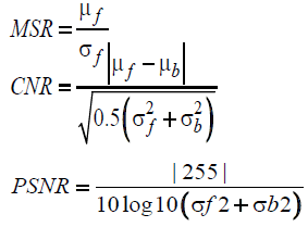
Quantitative measures of performance: We used meanto- standard-deviation ratio (MSR) contrast-to-noise ratio (CNR) [and peak signal-to-noise-ratio (PSNR) to compare the performance of different de-noising algorithms. The metrics MSR and CNR are defined as follows
Where μb and σ bare the mean and the standard deviation of the background region, while μf and σ f are the mean and the standard deviation of the foreground regions
From the above it can be observed that the proposed c compared to MSTD, KVSD and BM3D methods.
Where μb and σb are the mean and the standard deviation of the background region, while μ f and σ f are the mean and the standard deviation of the foreground regions.From the above it can be observed that there proposed approach has a better visual quality as compared to MSTD, KVSD, BM3D methods [21].
Conclusion
From the experimental results it is found that the proposed method outperforms the state of art algorithm in terms of objective & subjective visual quality for Speckle noise in Optic al tomography images with Reasonable computational cost compared to BM3D for retinal images that is due to additional pre-filtering step at initial stage.
References
- Drexler W. Ultrahigh-resolution optical coherence tomography J, Biomed. Opt. 2004; 9: 47- 74.
- Schmitt JM, Xiang SH, Yung KM. Speckle in optical coherence tomography. Journal of Biomedical Optics.1999;4:95-105.
- Adler DC, Koh TH, Fujimoto JG. Speckle reduction in optical coherence tomography images by use of a spatially adaptive wavelet filter. Optics Letters. 2004; 29:2878- 80.
- Lu Y.H, Tan SY, Yeo TS, et al. Adaptive filtering algorithms for sar speckle reduction. In Proc. IEEE 1996 International Geoscience and Remote Sensing Symposium.1996;67- 9.
- Fernandez DC, Salinas HM. Evaluation of a nonlinear diffusion process for segmentation and quantification of lesions in optical coherence tomography images. In Proc. Society of Photo Optical Engineers. 1834-43. San Jose, CA, International Journal of Computer Applications 2005;65:975-1887.
- Fernandez DC, Salinas HM, Puliafito CA. Automated detection of retinal layer structures on optical coherence tomography images. Opt. Express. 2005; 13: 200- 16.
- Chong B, Yong-Kai Z, Speckle reduction in optical coherence tomography images of human finger skin by wavelet modified BM3D filter. Original Research Article Optics Communications. 2013; 291: 461-9.
- Mashaly AS, AbdElkawy EEF, Mahmoud TA. Speckle noise reduction in SAR images using adaptive morphological filter, 10th Conference International on Intelligent Systems Design and Applications (ISDA), 2010.
- Starck JL, Fadili J, Murtagh F. The un-decimated wavelet decomposition and its reconstruction. IEEE Trans Image Process.2007;16:207-309.
- Gonzalez RC. Digital image processing. 2nd edition. Upper Saddle River, NJ: Pearson Education, 2004.
- Dabov K, Foi A, Katkovnik V, et al. Image de-noising by sparse 3D transform-domain collaborative filtering, IEEE Transactions on Image Processing 2007; 16: 2080-95.
- Kamalakshi N, ShanmukhaSwamy.“An efficient method of denoising” International Journal of Computer Applications in Engineering Sciences. 2013; 2100-40.
- Kamalakshi N, Naganna M, Shanmukhaswamy N, Modification of BM3D Algorithm for Representing Volumetric Data on medical images, International Journal of Computer Sciences and Engineering. 2013; 1: 2347-693.
- Kamalakshi N, ShanmukhaSwamy MN. An Approach For Modification Of Block Match Three Dimension De-Noising Algorithm With Pre-Filtering In Nonlocal Domain, International Journal of Innovative Science, Engineering ISSN 2015; 2: 2348-7968.
- Marc Lebrun. An Analysis and Implementation of the BM3D Image De-noising Method IPOL. 2012.
- Fang S, Li S, Nie Q, et al. Sparsely Based De-noising of Spectral Domain Optical Coherence Tomography Images Bio-Medical Optics Express. 2012; 3: 927-42.
- Marc L. An Implementation and detailed Analysis of the KSVD Image De-noising Method IPOL 2012.
- Chen Q, Sisternes L, Leng T, et al. Application of improved homogeneity similarity-based de-noising in optical coherence tomography retinal images. J Digit Imaging. 2015;1: 346-61.
- Buades A, Coll B, Morel JM. A Review of Image De-noising Algorithms, with a New One. Multiscale Modeling and Simulation. 2005; 2:490-530.
- Buades A, Coll B, Morel JM. Image De-noising Methods. A New Nonlocal Principle. SIAM Review. 2010;52: 113-47.
- Buades A, Coll B, and Morel J.M. Non-Local Means De-noising 2010.