Research Article - Biomedical Research (2017) Volume 28, Issue 8
Comparison of FLAIR brain MRI with different TEs (100, 165 ms) in detection of multiple sclerosis lesions
Maryam Razzaz*, Farnaz Fahimi, Fatemeh Meymanadinia and Narges Khanjani
Besat MRI Center, Kerman University of Medical Sciences, Kerman, Iran
Accepted date: January 9, 2017
Abstract
One of the most important parameters in Fluid-Attenuated Inversion Recovery (FLAIR) sequences MRI (FLAIR-MRI) is Echo Time (TE), so in this study we compare 100 ms and 165 ms echo time to diagnosis of Multiple Sclerosis (MS) lesions and use it for Flair-MRI protocol in MS patients. In this study we acquired FlAIR images at TE1=100 ms and TE2=165 ms with 1.5 Tesla (T) from 55 patients with MS. And then these images were studied to determine the number and location of MS lesions. Supratentorially and infratentorially, there were significant differences between the two sequences, in 100 ms echo time the number of lesions were 25.52 ± 20.69 (supratentorial), 0.58 ± 0.80 (infratentorial) and in 165 ms echo time 29.04 ± 20.69 (supratentorial), 0.67 ± 0.81 (infratentorial). (P Value=0.000). The presented results suggest that by increasing echo time of FlAIR-MRI diagnosis of MS lesions will be increased. So we can detect the lesions in early stages of the disease and also in follow-up studies.
Keywords
Multiple sclerosis, FLAIR-MRI, Echo time, Supratentorial, Infratentorial
Introduction
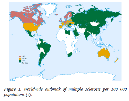
Multiple Sclerosis (MS) is a demyelinating disease of the Central Nervous System (CNS), mainly influences not only the white matter but also parts of the gray matter in the brain [1-4]. As a result, MS affects the brain and spinal cord and causes disabilities in young people. Early MS symptoms include weakness, tingling, numbness, double vision, blindness in one eye, muscle weakness, trouble with sensation, or trouble with coordination. Other signs are muscle stiffness, thinking problems, and urinary problems [5]. MS is conceptualized as a complex disease, in which several environmental elements and genetic factors act together to result disease [6]. In addition, it is believed that MS is an autoimmune disorder because of showing some similarity with other autoimmune disease, such as predominance of affected women like systemic lupus erythematosus, as an autoimmune disorder [7]; and manifestation with other autoimmune disease not only in affected individuals but also in their family members [8]. Depending on the region, the various risks of MS has been reported [9]. For instance, tropical areas are low prevalence for MS, while the disease is common in temperature areas (Figure 1).
Figure 1: Worldwide outbreak of multiple sclerosis per 100 000 populations [7].
Different diagnostic criteria, including early diagnostic [10] and later diagnostic [11] are used to determine MS in patients. Based on these different criteria, several diagnostic methods are used such as neurological examination, Cerebrospinal Fluid (CSF), neurophysiological studies and Magnetic Resonance Imaging (MRI). Among all these methods, MRI shows a great sensitive assessment for detection of subtle white matter lesions in the brain [12]. Young et al. demonstrated a better sensitivity of MRI in detection of MS lesions [13]. Besides, Jackson and colleagues reported the superior sensitivity for the detection of lesions by MRI in patients both with definite MS and with chronic progression. While, in the case of acute phase, a similar sensitivity was reported by CT scan and MRI [14]. MRI is established as a multi sequence protocol, such as T2-weighted, precontrast and postcontrast T1-weighted, and Fluid-Attenuated Inversion Recovery sequences (FLAIR) for diagnosis of MS [15,16]. FLAIR- MRI provides the highest sensitivity in the detection of lesions close to the CSF [17,18]. Also, FLAIR-MRI sequence inhibits the signal of the adjacent CSF hence exhibits the lesions of MS more apparent [19]. The most reliable method for radiological diagnosis of MS is the use of T1W, T2W and FLAIR sequences in MRI. Lesions are much better seen in T2W, and the accuracy of diagnosis increases adjust to the ependymal with FLAIR. Although, T1W is less sensitive, it has the ability to recognize of old lesions in the corpus callosum. Therefore, usage of contrast to determine new lesions through T1W would be helpful [20]. Hajnal et al. reported the use of FLAIR pulse sequences to suppress the signal from CSF, whereas heavy T2 weighting can be reached by long Echo Time (TE) [21]. Hence, one of the most important parameters in Flair-MRI is Echo Time (TE). So, in this study we compare 100 ms and 165 ms echo time with 1.5 Tesla to diagnosis of MS lesions and use it for Flair-MRI protocol in MS patients.
Patients and Methods
At first, fifty-five patients with clinically definite multiple sclerosis were assessed on Beasat MRI center of Kerman Medical Sciences University by a single neurologist who personally reviewed all available clinical data and contacted patients. 78.3% of patients were female and the rest, 21.7%, were male. Also, 8.7% of individuals had another disease along with their main disease (multiple sclerosis). While, 87% of the patients were receiving necessary treatment for MS. Nine patients were removed from this investigation because of tumefactive multiple sclerosis, in which the central nervous system has multiple demyelinating lesions with irregular characteristics in spite of standard Multiple Sclerosis (MS). Mean age of the 46 remaining patients were 32.52 ± 8.87 years old, and the average time from the diagnosis has been long past was 3.57 ± 3.04 years (Table 1).
| Number | Least | Most | Average | Standard deviation | |
|---|---|---|---|---|---|
| Age (year) | 46 | 16 | 56 | 32.5217 | 8.87378 |
| Duration of diagnosis (year) | 46 | 0 | 13 | 3.5733 | 3.04558 |
Table 1. Average age and duration of disease detection in samples.
In this center the studies were cross-sectional and each patient had an MRI exam with different sequences, including T1W, T2W and FLAIR-MRI with two various Echo Time (TE), 100 ms and 165 ms, for the comparison detection of supratentorial and infratentorial brain lesions in MS patients. After that, each stereotype of MRI was assessed to identify the exact number and location of old lesions and the new one, which enhanced through post-contrast T1W, related to MS by an experienced radiologist. Each stereotype of MRI was read randomly and done as a double blind experiment, in which the identity of those receiving a test treatment was concealed from both administrators and subjects until after the study was completed. Then, the result was encoded into SPSS software to compare.
Results
Considering the number of multiple sclerosis lesions that could be seen in two different Echo Time (TE) was performed by FLAIR-MRI and was described the experiment for testing the analytical predictions. A comparison of the average number of lesions detectability was performed for the supratentorial region at 100 ms and 165 ms echo time was 22.52 ± 20.29 and 29.04 ± 20.69 respectively (Table 2).
| Echo time | Number of lesions | Average | Standard deviation | P value |
|---|---|---|---|---|
| 100 ms | 1174 | 25.5217 | 18.37963 | 0.000 |
| 165 ms | 1336 | 29.0435 | 20.69348 | 0.000 |
Table 2. The mean number of lesions at different echo time in the supratentorial region.
Also, in infratentorial region the average number of MS lesions for 100 ms and 165 ms echo time was 0.58 ± 0.80 and 0.67 ± 0.81 subsequently (Table 3). As the data shown, the number of lesions in echo time of 165 ms was significantly more than the echo time of 100 ms (P Value=0.000).
| Echo time | Number of lesions | Average | Standard deviation | P value |
|---|---|---|---|---|
| 100 ms | 27 | 0.5870 | 0.80488 | 0.000 |
| 165 ms | 31 | 0.6739 | 0.81797 | 0.000 |
Table 3. The mean number of lesions at different echo time in the infratentorial region.
Discussion
The purpose of this study was to identify the optimal TE for FLAIR-MRI at 1.5 T assessing two different echo times to evaluate an appropriate diagnosis for MS lesions. Based on the data given by 100 and 165 ms TE, the best echo time for both infratentorial and supratentorial regions was 165 ms. In addition, some other assessments are also done in this field to find the optimum TE with more contrast and accuracy for FLAIR-MRI in detection of MS. One of the first study was taken into account by Raybrg et al. for the effect of selection of the echo time, and the echo time of 140 ms was introduced as a proper echo time that providing maximum contrast [22]. Pikus et al. claimed the improving detection of artificial Multiple Sclerosis (MS) lesions that were randomly distributed supraand infratentorially on simulated FLAIR-MRI obtained at different echo times [23]. In another study, Beckman et al. showed FLAIR-MRI with three different echo times of 100, 120 and 140 ms to evaluate the best one for detection of MS, and the most powerful identification was done at the echo time of 120 ms [24]. Besides, the stereotypes of FLAIR-MRI with echo times of 90 and 155 ms was investigated by Polman et al. which pointed that the more echo time, the more lesions in supratentorial region; although the number of false positive lesions would increase as well. While there was no difference for detection of infrantentorial lesions with these two echo times [25].
References
- Trapp BD, Peterson J, Ransohoff RM, Rudick R, Mörk S. Axonal transection in the lesions of multiple sclerosis. N Engl J Med 1998; 338: 278-285.
- Kidd D, Barkhof F, McConnell R, Algra PR, Allen IV. Cortical lesions in multiple sclerosis. Brain 1999; 122: 17-26.
- Ge Y, Grossman RI, Udupa JK, Babb JS, Kolson DL, McGowan JC. Magnetization transfer ratio histogram analysis of gray matter in relapsing-remitting multiple sclerosis. Am J Neuroradiol 2001; 22: 470-475.
- Geurts JJ, Bo L, Pouwels PJ, Castelijns JA, Polman CH, Barkhof F. Cortical lesions in multiple sclerosis: combined postmortem MR imaging and histopathology. Am J Neuroradiol 2005; 26: 572-577.
- Rejdak K, Jackson S, Giovannoni G. Multiple sclerosis: a practical overview for clinicians. Br Med Bull 2010; 95: 79-104.
- Marrie RA. Environmental risk factors in multiple sclerosis aetiology. Lancet Neurol 2004; 3: 709-718.
- Cooper GS, Stroehla BC. The epidemiology of autoimmune diseases. Autoimmun Rev 2003; 2: 119-125.
- Confavreux C, Hutchinson M, Hours MM, Cortinovis-Tourniaire P, Moreau T. Rate of pregnancy-related relapse in multiple sclerosis. Pregnancy in Multiple Sclerosis Group. N Engl J Med 1998; 339: 285-291.
- Pugliatti M, Sotgiu S, Rosati G. The worldwide prevalence of multiple sclerosis. Clin Neurol Neurosurg 2002; 104: 182-191.
- Schumacher GA, Beebe G, Kibler RF, Kurland LT, Kurtzke JF, McDowell F. Problems of experimental trials of therapy in multiple sclerosis: report by the panel on the evaluation of experimental trials of therapy in multiple sclerosis. Ann NY Acad Sci 1965; 122: 552-568.
- Poser CM, Paty DW, Scheinberg L, McDonald WI, Davis FA, Ebers GC. New diagnostic criteria for multiple sclerosis: guidelines for research protocols. Ann Neurol 1983; 13: 227-231.
- Wattjes MP, Lutterbey GG, Harzheim M, Gieseke J, Traber F, Klotz L. Higher sensitivity in the detection of inflammatory brain lesions in patients with clinically isolated syndromes suggestive of multiple sclerosis using high field MRI: an intraindividual comparison of 1.5 T with 3.0 T. Eur Radiol 2006; 16: 2067-2073.
- Young IR, Hall AS, Pallis CA, Legg NJ, Bydder GM. Nuclear magnetic resonance imaging of the brain in multiple sclerosis. Lancet 1981; 2: 1063-1066.
- Jackson J, Leake D, Schneiders N, Rolak L, Kelley G, Ford J. Magnetic resonance imaging in multiple sclerosis: results in 32 cases. Am J Neuroradiol 1985; 6: 171-176.
- Simon J, Li D, Traboulsee A, Coyle P, Arnold D, Barkhof F. Standardized MR imaging protocol for multiple sclerosis: Consortium of MS Centers consensus guidelines. Am J Neuroradiol 2006; 27: 455-461.
- Filippi M, Falini A, Arnold DL, Fazekas F, Gonen O, Simon JH. Magnetic resonance techniques for the in vivo assessment of multiple sclerosis pathology: consensus report of the white matter study group. J Magn Reson Imag 2005; 21: 669-675.
- Frohman E, Zhang H, Kramer P, Fleckenstein J, Hawker K, Racke M. MRI characteristics of the MLF in MS patients with chronic internuclear ophthalmoparesis. Neurology 2001; 57: 762-768.
- Gawne-Cain M, ORiordan J, Thompson A, Moseley I, Miller D. Multiple sclerosis lesion detection in the brain: a comparison of fast fluid-attenuated inversion recovery and conventional T2-weighted dual spin echo. Neurology 1997; 49: 364-370.
- Hashemi RH, Bradley WG, Chen DY, Jordan JE, Queralt JA, Cheng AE. Suspected multiple sclerosis: MR imaging with a thin-section fast FLAIR pulse sequence. Radiology 1995; 196: 505-510.
- Karolyi DR. CT and MRI of the Whole Body. Acad Radiol 2009; 16: 1451.
- Hajnal JV, De Coene B, Lewis PD, Baudouin CJ, Cowan FM, Pennock JM. High signal regions in normal white matter shown by heavily T2-weighted CSF nulled IR sequences. J Comp Assist Tomogr 1992; 16: 506-513.
- Rydberg JN, Riederer SJ, Rydberg CH, Jack CR. Contrast optimization of fluid-attenuated inversion recovery (FLAIR) imaging. Magn Reson Med 1995; 34: 868-877.
- Pikus L, Woo JH, Wolf RL, Herskovits EH, Moonis G, Jawad AF. Artificial multiple sclerosis lesions on simulated FLAIR Brain MR images: echo time and observer performance in detection. Radiology 2006; 239: 238-245.
- Bachmann R, Reilmann R, Schwindt W, Kugel H, Heindel W. FLAIR imaging for multiple sclerosis: a comparative MR study at 1.5 and 3.0 Tesla. Eur Radiol 2006; 16: 915-921.
- Polman CH, Reingold SC, Banwell B, Clanet M, Cohen JA. Diagnostic criteria for multiple sclerosis: 2010 revisions to the McDonald criteria. Ann Neurol 2011; 69: 292-302.
