Image Article - Journal of Gastroenterology and Digestive Diseases (2023) Volume 8, Issue 6
COLONIC LEIOMYOMA: AN UNCOMMON OCCURRENCE
Sahar Hamza1*, Ines Chelly2, Lamia Kallel11Gastro-enterology Department, Mahmoud El Matri Hospital, University of Tunis El Manar, Tunisia
2Anatomopathology Department, University of Tunis EI Manar, Tunis, Tunisia
- *Corresponding Author:
- Sahar Hamza
Gastro-enterology Department
Mahmoud El Matri Hospital
University of Tunis El Manar, Tunis, Tunisia
E-mail: Sahar.hamza137@gmail.com
Received: 29-Dec-2023, Manuscript No. JGDD-23-123889; Editor assigned: 03-Jan-2024, Pre QC No. JGDD-23-123889 (PQ); Reviewed: 15-Jan-2024, QC No. JGDD-23-123889;
Revised: 19-Jan-2024, Manuscript No. JGDD-23-123889 (R); Published: 25- Jan-2024, DOI: 10.35841/ jgdd -8.6.180
Citation: Hamza S, Chelly I, Kallel L. Colonic leiomyoma: An uncommon occurrence. J Gastroenterol Dig Dis. 2023;8(6):180
A 53-year-old, previously healthy patient, presented to the gastroenterology department with a 6-month history of abdominal pain, and minimal bright red blood per rectum. She denied having other symptoms including constipation, diarrhea, or weight loss. Physical exam and laboratory testing were non-contributory. Proctoscopy showed internal haemorroids but there was no signs of active bleeding.
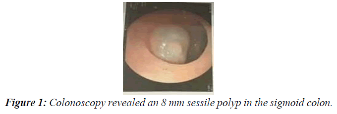
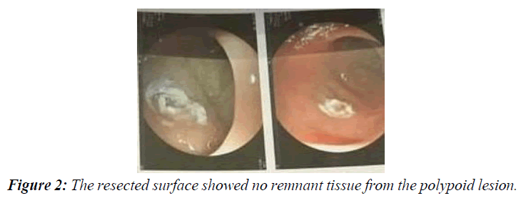
Colonoscopy revealed an 8 mm sessile polyp in the sigmoid colon (Figure 1). Submucosal saline-adrenaline injection was done to lift the lesion for polypectomy. The polyp was removed by hot snare polypectomy and sent for histopathology. The resected surface showed no remnant tissue from the polypoid lesion (Figure 2).
Histologic examination revealed an architecturally preserved colonic mucosa lifted by a fuso-cellular proliferation of smooth muscle margins cells organized in tangled bundles, with no mitosis. Immunohistological findings were negative for Cluster of Differentiation (CD) 117 and DOG1, ruling out gastrointestinal stromal tumors (Figure 3).
These findings were consistent with a diagnosis of submucosal leiomyoma. No bleeding or perforation was noted after the polypectomy.


