Clinical Image - Journal of Gastroenterology and Digestive Diseases (2017) Volume 2, Issue 3
Atypical endoscopic finding for chronic Helicobacter pylori gastritis.
Fady Maher Wadea*
Faculty of Medicine, Department of Internal Medicine, Gastroenterology and Hepatology division, Zagazig University, Zagazig, Egypt
- *Corresponding Author:
- Fady Maher Wadea
Faculty of Medicine Department of Internal Medicine, Gastroenterology and Hepatology division Zagazig University Zagazig Egypt
Tel: +20 55 2364612
E-mail: fadymaher41@yahoo.coms
Accepted Date: December 22, 2017
Citation: Wadea FM, Atypical endoscopic finding for chronic Helicobacter pylori gastritis. J Gastroenterol Dig Dis. 2017;2(3):20.
Abstract
Female patient 60-year-old age with no history of previous medical diseases apart from HCV chronic hepatitis, complain of persistent epigastric pain and recurrent vomiting, lack of appetite and loss of weight in previous two months. No haematemesis or melena is present nor dysphagia. Patient sought medical advice and take different types of P.P.I preparations without improvement.
Clinical Image
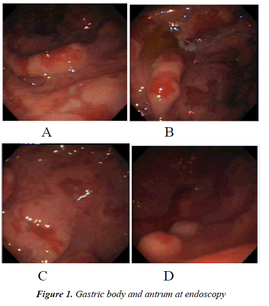
Female patient 60-year-old age with no history of previous medical diseases apart from HCV chronic hepatitis, complain of persistent epigastric pain and recurrent vomiting, lack of appetite and loss of weight in previous two months. No haematemesis or melena is present nor dysphagia. Patient sought medical advice and take different types of P.P.I preparations without improvement. Patient ultrasonography was normal, CT abdomen with contrast shows thickened gastric mucosa suspected with gastric lymphoma for upper GIT endoscopy and biopsy. Endoscopy shows pangastritis with diffuse multiple superficial ulceration and inflammatory polyps and highly congested edematous mucosa and excessive biliary reflux (Figures 1A-1D). Duodenal mucosa shows mild duodenitis; multiple biopsies were taken for histopathological examination. Biopsy was examined by at least 3 pathologists at different times with intra observer agreement shows that gastric mucosa shows moderate inflammatory exudates rich in lymphocytes, plasma cells and neutrophils with curvilinear bacilli in glands lumen consistent with Helicobacter pylori organism associated with inflammatory polyp. No evidence of intestinal metaplasia, dysplasia or malignancy or lymphoma. The picture was consistent with chronic H. pylori infection of moderate activity. The endoscopic picture is included which was different from typical mucosal modularity and erythematous mottling seen frequently at endoscopy. Eradication therapy for H.pylori was given to patient and repeated endoscopy and biopsy was done that confirmed absence of malignancy or lymphoma cells. Patient's symptoms was improved apart from periods of epigastric pain that improve with P.P.I use, however endoscopic picture shows partial improvment. Patient was advised for regular follow up.
Conflict of Interest
None to declare.
