Research Article - Biomedical Research (2016) Volume 27, Issue 4
Asymptomatic bilateral calcified stylohyoid ligaments detection by panoramic radiography and cone beam computerized tomography
Ramon Fuentes1*, Diego Saravia2,3, Ivonne Garay4, Nicolas Ernesto Ottone1,5
1Research Centre in Dental Science (CICO), Dental School, Universidad de La Frontera, Temuco, Chile
2Universidad Adventista de Chile, Chillan, Chile
3Master Program in Dentistry, Dental School, Universidad de La Frontera, Temuco, Chile
4Oral and Maxillofacial Imaging Unit, Dental School, Universidad de La Frontera, Temuco, Chile
5Doctoral Program in Morphological Sciences, Medicine School, Universidad de La Frontera, Temuco, Chile
- *Corresponding Author:
- Prof. Dr. Ramon Fuentes
CICO Centro de Investigacion en Ciencias Odontologicas
Universidad de La Frontera
Manuel Montt 112, Casilla 54-D
Temuco, Chile
Accepted date: April 28, 2016
Abstract
Stylohyoid ligaments calcified are found incidentally through imaging. Morphological characteristics can determine the development of different pathological compression levels of the head and neck. We report a case of an asymptomatic bilateral calcification of stylohyoid ligaments visualized by panoramic radiography and cone beam computerized tomography reconstruction. The correct description and visualization of this anomaly is important to carry out an accurate diagnosis and appropriate treatment in patients who present it.
Keywords
Stylohyoid ligament calcification, Bilateral, Panoramic radiography, Cone beam Computerized tomography.
Introduction
The styloid process is a thin cylindrical bony projection, measuring 25 mm on average. Its origin is in the tympanic portion of the temporal bone. An elongated styloid process and calcified styloid ligaments may be part of a condition known as Eagle Syndrome or Stylohyoid Syndrome [1-4]. The term of elongated styloid process was described by Eagle in 1973 [5], and the elongated styloid process related to a stylohyoid ligament ossification was described by Pietro Marchetti in 1652 [5]. Eagle considered as an abnormal styloid process when it was longer than 25 millimetres in adults, instead styloid process normal size varies significantly between 20 and 30 millimetres [6,7]. It has been reported that the population affected by an elongated styloid process is about 4%, however, from this percentage, only 4% have symptoms [8-10]. It has been reported as well, that this condition is more prevalent in women than in men [11,12]. This syndrome includes a foreign body sensation in the throat, sore, dysphagia and sometimes pain in the neck or in the face when exists compression of cranial nerves due to the characteristics of the calcified stylohyoid ligament and elongated styloid process [13]. This syndrome may also present cerebrovascular commitment which is manifested with headaches, syncope and transitory loss of vision when the internal carotid artery is affected [14]. Eagle classified this syndrome into two: classic type and carotid artery type. The first may presents symptoms of foreign body sensation, referred ear pain and dysphagia. The second one may presents symptoms such as headaches and neurological symptoms, and pain in temporal and maxillary branches [15,16]. This syndrome has been reported on twins showing the same pattern [17], and this syndrome is uncommonly suspected in clinical practice [18].
Case Report
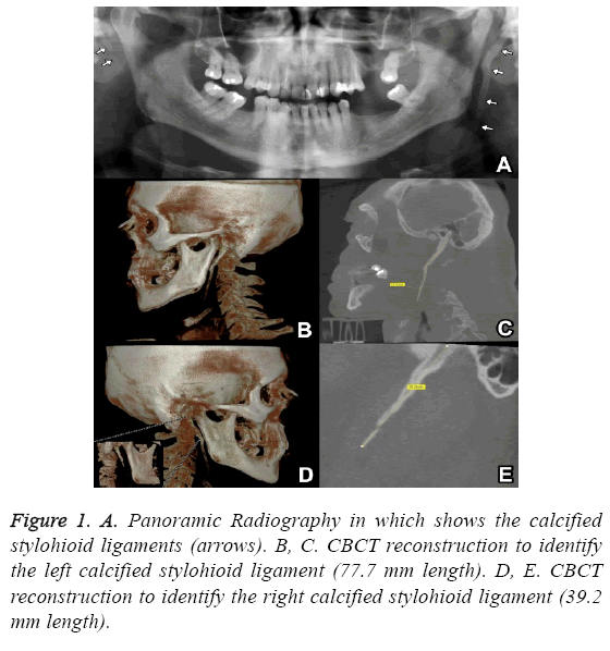
A male patient, 55 years old, with no relevant medical history and without symptomatology from any dental pathology was studied. He presents an osteofito in the right mandibular condyle, and considerable ossification of the left styloid process. A panoramic radiograph is initially taken, when this condition was found. The patient looks for treatment of teeth 1.2 and 2.5 with fixed singular prosthesis (Figure 1A). A cone beam computerized tomography (CBCT) examination was then performed showing ossification of both ligaments (Figures 1B-1E). The left stylohioid ligament, in the measurement of the proximal section, is continuous and with a pseudoarticulated aspect (Figure 1B). On the right side, the stylohioid ligament is discontinuous (Figure 1D). Degenerative bone remodelling process is also observed in both condyles, right condyle is greater in size. The ossified right ligament has a length of 39.2 mm (Figure 1E). The left ligament is longer (77.7 mm) (Figure 1C).
Discussion
An elongated styloid process is a calcification of soft tissue that can be identified in panoramic radiographs. Is not commonly treated by surgery, because most of the time it doesn’t imply any symptoms for the patients. Sometimes, this condition has been considered as an anatomical variation. Normal length of the styloid process varies between 25 and 30 mm, and above 30 mm is considered as elongated [10,19-22]. The embryological origin of this complex is in the second bronchial arch. During its development, the cartilage portion of the second arch extends itself to each side from the otic capsule to the median line, where the styloid process, stylohyoid ligament, lower cusp and the cranial border of the hyoid bone are formed. Then, the cartilage portion of the second arch joins the styloid process with the hyoid. As a consequence of the ossification of the styloid process, the styloid ligament could extend this ossification originating elongated styloid process [23]. In some cases the styloid process and the hyoid can remain adjoined [24]. Calcified stylohyoid ligaments are thought to be the result of traumatic scarring or posttonsillectomy. Has been reported that a trauma of the cervical and throat region, especially after tonsil surgery, can stimulates styloid process growth [14]. It is still controversial the relation between trauma history and calcification of stylohyoid complex, due to in many cases with this condition there is not previous trauma. It has been suggested that persistence of mesenchyme elements [25], bone tissue growth, endocrine disorders in women with menopause, mechanical stress, could result in a calcified hyperplasia of the stylohyoid ligament. Okabe et al. found a significant correlation between the size of the calcification of stylohyoid complex and serum calcium concentrations along with bone density [26]. MacDonald- Jankowski reported significative differences in stylohyoid ligament morphology between patients from London and Hong Kong with can indicate that may exist genetic influences [27]. The bilateral ossification of the stylohyoid ligamen is not common and has a low frequency (4%). This condition can be asymptomatic as described in this case and by others authors before [8,9].
Conclusion
It may be difficult to detect this anatomical variation using a conventional 2D panoramic radiography. Although in this case the radiography allowed identifying the ossified styloid process, we consider that CBCT technology is very important exam for the diagnostic of this anatomical conditions which makes possible to perform reconstructions in three dimensions, multi cut images and precisely identification of morphological characteristics [24]. In patients with symptoms due to ossified stylohyoid ligament, the CBCT exam could help to perform an accurate diagnosis, because as symptoms are diverse and not specific, patients usually look for treatment in different areas such as otolaryngology, neurology, psychiatry and dentistry.
References
- Winkler S, Sammartino FJ Sr, Sammartino FJ Jr, Monari JH. Stylohyoid Syndrome. Report Of A Case. Oral Surg Oral Med Oral Pathol 1981; 51: 215-217.
- Catelani C, Cudia G. Stylalgia Or Eagle Syndrome. Report Of A Case. Dent Cadmos 1989; 57: 70-74.
- Babad MS. Eagle's Syndrome Caused By Traumatic Fracture Of A Mineralized Stylohyoid Ligament-Literature Review And A Case Report. Cranio 1995; 13: 188-192.
- Feldman VB. Eagle’s Syndrome: A Case Of Symptomatic Calcification Of The Stylohyoid Ligaments. J Can Chiropr Assoc 2003; 47: 21-27.
- Eagle WW. Elongated Styloid Processes: Report Of Two Cases. Arch Otolaryngol 1937; 25: 584-587.
- Ilguy M, Ilguy D, Guler N, Bayirli G. Incidence Of The Type And Calcification Patterns In Patients With Elongated Styloid Process. J Int Med Res 2005; 33: 96-102.
- Monsour PA, Young WG. Variability Of The Styloid Process And Stylohyoid Ligament In Panoramic Radiographs. Oral Surg Oral Med Oral Pathol 1986; 61:522-526.
- Johnson GM, Rosdy NM, Horton SJ. Manual Therapy Assessment Findings In Patients Diagnosed With Eagle's Syndrome: A Case Series. Man Ther 2011; 16: 199-202.
- Yavuz H, Caylakli F, Erkan AN, Ozluoglu LN. Modified Intraoral Approach For Removal Of An Elongated Styloid Process. J Otolaryngol Head Neck Surg 2011; 40: 86-90.
- Natsis K, Repousi E, Noussios G, Papathanasiou E, Apostolidis S, Piagkou M. The Styloid Process In A Greek Population: An Anatomical Study With Clinical Implications. Anat Sci Int 2015; 90: 67-74.
- Fuentes FR, Oporto VG, Garay CI, Bustos Ml, Silva MH, Flores FH. Styloid Process In The Panoramics Radiographic Sample Of Temuco-Chile City. Int J Morphol 2007; 25: 729-733.
- Garay I, Olate S. Ossification Of The Stylohyoid Ligament In 3028 Digital Panoramic Radiographs. Int J Morphol 2013; 31: 31-3
- Haynes MJ, Vincent K, Fischhoff C, Bremner AP, Lanlo O, Hankey GJ. Assessing The Risk Of Stroke From Neck Manipulation: A Systematic Review. Int J Clin Pract 2012; 66: 940-947.
- Fusco DJ, Asteraki S, Spetzler RF. Eagle's Syndrome: Embryology, Anatomy, And Clinical Management. Acta Neurochir 2012; 154: 1119-1126.
- Colby CC, Del Gaudio JM. Stylohyoid Complex Syndrome: A New Diagnostic Classification. Arch Otolaryngol Head Neck Surg 2011; 137: 248-252.
- Costantinides F, Vidoni G, Bodin C, Di Lenarda R. Eagle's Syndrome: Signs And Symptoms. Cranio 2013; 31: 56-60.
- Kim JE, Min JH, Park HR, Choi BR, Choi JW, Huh KH. Severe Calcified Stylohyoid Complex In Twins: A Case Report. Imaging Sci Dent 2012; 42: 95-97.
- Fini G, Gasparini G, Filippini F, Becelli R, Marcotullio D. The Long Styloid Process Syndrome Or Eagle's Syndrome. J Craniomaxillofac Surg 2000; 28: 123-127.
- Kaufman SM, Elzay RP, Irish EF. Styloid Process Variation. Radiologic And Clinical Study. Arch Otolaryngol 1970; 91: 460-463.
- Keur JJ, Campbell JP, Mccarthy JF, Ralph WJ. The Clinical Significance Of The Elongated Styloid Process. Oral Surg Oral Med Oral Pathol 1986; 61: 399-404.
- Sokler K, Sandev S. New Classification Of The Styloid Process Length--Clinical Application On The Biological Base. Coll Antropol 2001; 25: 627-632.
- Scaf G, Freitas DQ, Loffredo LC. Diagnostic Reproducibility Of The Elongated Styloid Process. J Appl Oral Sci 2003; 11: 120-124.
- Moore Kl, Persaud T. The Developmental Human Clinically Oriented Embryology. Philadelphia W.B. Saunders Company 1998.
- Yagci AB, Kiroglu Y, Ozdemir B, Kara CO. Three-Dimensional Computed Tomography Of A Complete Stylohyoid Ossification With Articulation. Surg Radiol Anat 2008; 30:167-169.
- Rechtweg JS, Wax MK. Eagle's Syndrome: A Review. Am J Otolaryngol 1998; 19:316-321.
- Okabe S, Morimoto Y, Ansai T, Yamada K, Tanaka T, Awano S, Kito S, Takata Y, Takehara T, Ohba T. Clinical Significance And Variation Of The Advanced Calcified Stylohyoid Complex Detected By Panoramic Radiographs Among 80-Year-Old Subjects. Dentomaxillofac Radiol 2006; 35:191-199.
- Macdonald-Jankowski DS. Calcification Of The Stylohyoid Complex In Londoners And Hong Kong Chinese. Dentomaxillofac Radiol 2001; 30:35-39.
