- Biomedical Research (2016) Volume 27, Issue 2
An automatic clinical decision support system intended for MRI Brain images.
| Josephine Sutha V1*, Latha P2 1Sardar Raja College of Engineering, Tirunelveli, India. 2Government College of Engineering, Tirunelveli, India. |
| Corresponding Author: Josephine Sutha V Sardar Raja College of Engineering Tirunelveli India |
| Accepted: November 26, 2015 |
Abstract
Interpretation error and accuracy rate are the focal issues in traditional magnetic resonance image (MRI) classification. In order to decipher this issue accurate automatic detection and classification of images with prior knowledge is projected in this research paper. The principal objective of this research work is to form a classification system that should not be suffered by misclassification results so that it can be exaggerated up to countless years. Keep the requirements in attention, a hybrid technique for automatic classification of MR images with shift-invariant discrete wavelet transform (SIDWT) and adaptive neuro-fuzzy inference system (ANFIS) is advocated in this paper. Two different types of tumour explicitly glioma and Meningioma have been given considerable concentration. Feature extraction and classification are the two main stages in this work. In the first stage, five texture features, Contrast, Correlation, Energy, Homogeneity and Entropy are extracted using Gray Level Co-occurrence Matrix (GLCM). In the second stage, an ANFIS classifier is anticipated. The system was established for proficient in classification with the accuracy rate of 99.8%.Fast and accurate results in a reduced time are the main advantages of the suggested method.
Keywords |
||||||||||
| Tumour, Shift-Invariant discrete wavelet Transform, Adaptive Neuro-Fuzzy Inference System, Feature extraction. | ||||||||||
Introduction |
||||||||||
| Medical imaging is the run-through and knack of creating visual representations of the interior of a body for indisputable scrutiny and medical intrusion. Medical imaging launches a collection of normal anatomy and physiology to make it possible to categorize the abnormalities. Magnetic resonance imaging (MRI) is one of the medical imaging techniques customs in radiology to review the anatomy and the function of the body both in health and disease. Brain tumour, the frightful illness occurs in the abnormal cell formation in the brain. A brain tumour may be identified by MRI scans. Malignant or cancerous tumours and benign or harmless tumours are the two kinds of tumours form in brain [1]. All types of brain tumours could make indications, which vary depending on the part of the brain involved. Indications incorporate headaches, seizures, vision going delinquent, retching, and emotional vagaries [2]. | ||||||||||
| The purpose of the classification is to use the information contained in magnetic resonance (MR) images for quantitative applications [3]. Operator-assisted classification schemes are non-reproducible, and also not practical for the hefty amounts of data required for a meaningful statistical analysis. Numerous types of computerized investigation can be used to extract information from three-dimensional (3-D) MRI records of the human head [4]. Amongst, Manual or semi-automatic classification performed by the trained experts is labour- intensive for the quantitative analysis of MRI data. | ||||||||||
| Former knowledge can be executed in different ways. Selective methodologies have been acknowledged as shown by Arijita et al. [5], Bhawna and Shamik [6], Daljit and Kamaljeet [7] as well as Josephine and Latha (2014) for the classification of MR images in recent years. In studies by Kulhalli et al., Principal component analysis (PCA) with probabilistic neural network (PNN) is used for Primary Level Classification of Brain Tumour. Subsequently, the tested Magnetic Resonance Image (MRI) of brain is categorized as benign or malignant. In studies by Ramteke et al., Automatic medical image classification and abnormality detection using K-nearest neighbour is suggested. At this juncture, the input CT brain image has been classified as Normal or Abnormal. But the Classification rate is not equal to the estimated level. However, fine-tuned classification is highly required. Besides, certain Classification approaches [10] lack efficient tuning. Hence to enhance and to progress the proficiency of above said methods, it is vital to have a system deploying level set method with SIDWT. | ||||||||||
| Around the Globe, there are no widely accepted schemes for automatic and reliable tumour detection. Since the interpretation error is high [11], there is an abundant need and interest for automatic classification systems. Interpretation error is an error which occurs while doctors do the tumor evaluation process manually. It may be avoided using the proposed method.The classification system comprises of database that contains predefined patterns which compares with detected object to classify into proper category. In an automatic medical decision system, the medical staffs match the given set of test MR image with the pre-existing tumour images. As a result, counteractive solution can easily be recognized without the consultant opinion of any expert. This saves the life of the patients in advance. | ||||||||||
Projected Method |
||||||||||
| The projected automatic classification system consists of two different steps, feature extraction by gray level co-occurrence matrix, and classification by ANFIS. Data base collected from the scan centres includes both normal and abnormal brain images. At first brain images are transformed to gray scale image. Subsequently, SIDWT is applied to get different coefficients. Afterwards, five texture features contrast, correlation, energy, homogeneity and entropy of the MRI brain images are extracted through gray level co- occurrence matrix. An example texture features values of five different MR Images have been illustrated in Table 1. Finally, ANFIS classifier is employed to identify whether the given query image falls in ‘Normal’ or ‘Abnormal’ sets. When the result is normal, the image is concluded as Normal. Else, the outcome is known as ‘Benign’ or ‘Malignant’. If ‘Benign’, the result will be clinched, otherwise, Level set method will be employed to identify whether the stage of tumour is ‘Meningioma’ or ‘Glioma’ Figure 3. Level set method [12] is an arithmetical procedure for trailing interfaces and shapes of the tumour. Summary of the suggested work is exposed in Figure 1. | ||||||||||
Data base |
||||||||||
| The presented classification work is implemented with the data base placid from the scan centres of Tirunelveli, the southern part of India. A set of 970 MR Images were considered as the database. They were separated as: 670 for training in which 70 are Normal, 300 are Benign and 300 are Malignant and 300 for testing in which 100 are Benign, 120 are Malignant and 80 are absolutely Normal. All are T2-weighted axial plane images with 256 × 256 in-plane resolution. | ||||||||||
Shift-invariant DWT |
||||||||||
| The discrete wavelet transform (DWT) has been taken as a tool for feature extraction and classification problems [13]. The ability to localize structures of an image with good resolution can also be done by discrete wavelet transform in an effective manner. The outcome of this representation is unique in which imperative structures resembling edges and details are quantified resourcefully by means of less number of coefficients. As DWT is shift- variant, any texture metrics towed out from the wavelet coefficients too are shift-variant [14]. This reduces the classification performance of the system. To conflict this concept in medical image classification, a shift-invariant DWT (SIDWT) is exploited to make sure that merely translation invariant features are extracted. | ||||||||||
Feature extraction |
||||||||||
| Feature Extraction has been done by gray-level co- occurrence matrix. This is a statistical method to scrutinize texture values that considers the spatial relationship of pixels [15].This is also known as the gray-level spatial dependence matrix. Gray Level Co-occurrence Matrix (GLCM) of an image is calculated by examining the repeated pairs of pixel with its exact values. GLCM texture measures are classified as: First order texture measures, second order measures and higher order texture measures [16]. In the proposed method, the following features Contrast, Correlation, Energy, Homogeneity and Entropy are calculated. | ||||||||||
| Contrast: Contrast is a degree of local variation in the image. It presents the degree of the legibility of the image. | ||||||||||
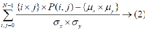
| Correlation: Correlation is the degree of gray tone undeviating dependencies in the image. | ||||||||||
 |
||||||||||
| Energy: Energy is a degree of the homogeneity of the image. | ||||||||||
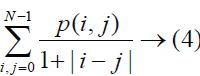
| Homogeneity: Homogeneity is the measure of the resemblance of gray scale levels athwart the image. | ||||||||||
 |
||||||||||
| Entropy: Entropy expresses the non-uniformity of an image grounded on probabilities of co-occurrence values. | ||||||||||
Adaptive Neuro-Fuzzy Inference System (ANFIS) |
||||||||||
| The proposed ANFIS classifier is the combined structure of neural network classifier and fuzzy classifier [17]. ANFIS has been taken as a universal estimator as its inference system corresponds to a set of fuzzy IF-THEN rules which have the learning capability for the nonlinear approximations. ANFIS is fundamentally a graphical network representation of Sugeno-type fuzzy systems endowed with the neural learning capabilities. The basic structure is represented in Figure 2. ANFIS architecture includes five layers with different nodes. The first and the fourth layer consist of flexible nodes while the remaining layers entail of predetermined nodes. Takagi-Sugeno method was introduced in 1985 and is computationally proficient that works fine with optimization and adaptive techniques, so that it can be applied in a variety of applications [18]. The Main motivation of Takagi-Sugeno is to shrink the number of rules essential for the system [19-25]. | ||||||||||
| A normal Sugeno type of fuzzy system use the subsequent rule base | ||||||||||
| 1. If x is A1 and y is B1, then |
||||||||||
| 2. If x is A2 and y is B2, then |
||||||||||
| Here x and y are the inputs, Ai and Bi are the fuzzy sets. fi is the output applied within the fuzzy area précised by the fuzzy rule. c11, c12, c10, c21, c22 and c20 are the proposed parameters, dogged during the training procedure. Let the fuzzy set membership functions are Ai, Bi, i=1, 2, be, μAi(x ) μBi( y) | ||||||||||
| In assessing the rules, select product for T-norm (logical AND). | ||||||||||
| 1. Evaluating the rule premises results in | ||||||||||
| 2. Evaluating the implication and the rule consequences gives | ||||||||||
 |
||||||||||
| ANFIS must be trained already so that it can get the practice as a classifier. To train the ANFIS, the input and output constraints of the membership functions need to be allotted. The training procedure adapts all the variable parameters of adaptive layers. Figure 4 shows the proposed diagram of a MRI Classifier. When the training got over, the testing data is used to test the accuracy of the ANFIS classifier. When the parameters of the membership function are modified, classification accuracy could be improved in accordance with the expected output. | ||||||||||
Results and Discussion |
||||||||||
| The ANFIS system is initialized for 300 iterations and the error tolerance value is 0.0001. Once the criterion is achieved, the network is supposed to be stabilized. The step size for parameter adaptation is 0.01. Also the input membership function has been taken as a bell curve type. Total data set was completely separated into two-one is the training data set and the other is testing data set. To train the ANFIS, training data set was applied, and to verify the accuracy of the proposed classifier, testing data set was applied. Besides, performance of the system has been evaluated according to the five measures: accuracy, sensitivity, specificity, precision and recall. These measures can be calculated by means of four options to be exact True Positive (TP), True Negative (TN), False Positive (FP) and False Negative (FN). | ||||||||||
| Where, | ||||||||||
| True Positive (TP): Correctly classified positive cases. | ||||||||||
| True Negative (TN): Correctly classified negative cases. | ||||||||||
| False Positive (FP): Wrongly classified negative cases. | ||||||||||
| False Negative (FN): Wrongly classified positive cases | ||||||||||
| Precision is a gauge of exactness, while Recall is a measure of fullness. Arithmetical classification task includes Precision as a class which is defined as the number of true positives divided by the total number of images categorized as fitting to the positive class. Recall is well-defined by the number of true positives with the total number of images that truly fit to the positive class. The values of different measures for the proposed method are given as below: | ||||||||||
| Accuracy = 99.8%, | ||||||||||
| Specificity = 98.50% and | ||||||||||
| Sensitivity = 100% | ||||||||||
| The highest classification accuracy for the testing Image was achieved with 100 training epochs. The classification accuracy was 99.79%. Table 2 shows the overall ANFIS performance Evaluation. Moreover, a set of five existing systems with different algorithms were taken for the accuracy enactment. Table 3 shows the performance of the system in terms of accuracy and execution time. From Table 3 it is observed that the average time taken to find the decision of the test image is 0.08 seconds. This protects the lifespan of patients in advance without wasting abundant time. As the proposed classifier is joined with the two techniques namely fuzzy classification and neural networks classification, it trounces the negative aspects of the individual classifier. Still, it has a drawback for its performance when applied for Large input data if the parameters not chosen suitably. That could be avoided by choosing suitable parameters. | ||||||||||
| Envisage the recital of the classifier can be done by means of receiver operating characteristics (ROC) graphs (3). ROC curve has been plotted linking the true positive rate (TPR) with the false positive rate (FPR). The TPR indicates the number of exact positive results come from all positive samples which are on hand all through the experiment. In contrast, FPR signifies the number of incorrect positive results comes from all negative samples which are on hand all through the experiment. Figure 5 shows Normal, Benign and Malignant outputs for a proposed classifier. | ||||||||||
Conclusion |
||||||||||
| Brain tumour causes bereavement. Many techniques are used to sense the tumour as early as possible because early detection may cure this disease. Medical Imaging provides many techniques for the identification of the tumour. In the proposed work, solutions have been suggested for the problems related with Interpretation error and Accuracy rate allied in automatic finding of MRI brain abnormalities. The experimental outcomes show that the proposed work gives less convergence time with good accuracy, sensitivity and specificity for the classification of MR brain abnormalities. Besides, the area under curve (AUC) for the proposed system has been calculated as 0.98. Hence, the proposed system has been shown with good accuracy. It also surmounts the drawbacks of artificial neural network classifier and fuzzy classifier by putting together. | ||||||||||
Tables at a glance |
||||||||||
|
||||||||||
Figures at a glance |
||||||||||
|
||||||||||
References |
||||||||||
|


