- Biomedical Research (2014) Volume 25, Issue 3
The morfometrical and morphological analysis of the acromion with Multidetector Computerized Tomography.
Musa ACAR1, Tuba ŞİMŞEK1, Mahinur ULUSOY2, İsmail ZARARSIZ2, Serpil ACAR3 and Duran EFE41Mevlana University, Department of Physical Therapy and Rehabilitation 42003 Konya TURKEY
2Mevlana Üniversity Faculty of Medicine Department of Anatomy
3Konya Numune Hospital Department of Physical Therapy and Rehabilitation
4Mevlana Üniversity Faculty of Medicine Department of Radiology
- *Corresponding Author:
- Musa ACAR
Mevlana University, Department of Physical
Therapy and Rehabilitation, 42003 Konya TURKEY
Accepted date: April 29 2014
Abstract
Subacromial compression is a clinical condition which appears due to compression of supraspinatus tendon in the subacromial space and causes severe shoulder pain. If subacromial compressions are not treated, this results with rotator cuff ruptures. One of the most important factors for appearance of subacromial compression syndrome and rotaror cuff ruptures is the morphology of acromion. We planned this study as our data would help for differential diagnosis and orthopedics during surgical treatment. This study was conducted on 73 patients (30 females-43 males). For morphometric assessment of the acromion, acromion width (AW), acromion length (AL) and the distance between the acromion and coracoid process (A-CP) were measured. We found in 55 (37%) subjects with flat acromion which is known as Type I, in 71 (48.7%) subjects with curved acromion which is known Type II and in 20 (13.7%) subjects with hooked acromion which is known as Type III. To know anatomic details and variations of the region is important for diagnosis and treatment. Furthermore, we think that our study is useful for creation of statistical and reference information of Turkish community.
Keywords
acromion, impingement, shoulder, variation
Introduction
Scapula has three processes including spina scapula, coracoid process and acromion. Acromion is on the posterior side of the scapula as an widening and flattening extension of the osseous process called spina scapulae to the lateral side. The coronoid shaped process on the superior side of glenoideal cavity is called corcoid process [1]. The area between acromion, coracoid process and coracoacromial ligament which extends among these two processes is called coracoacromial space [2].
Subacromial compression is a clinical condition which appears due to compression of supraspinatus tendon in the subacromial space and causes severe shoulder pain [1]. This syndrome narrows operation area of the rotator cuff and causes shoulder pain and movement restriction [3].
If subacromial compressions are not treated, this results with rotator cuff ruptures. One of the most important factors for appearance of subacromial compression syndrome and rotaror cuff ruptures is the morphology of acromion [4,5]. Three acromion types have been defined in the studies performed in the literature and a strong relationship has been detected with type II acromion, subacromial compression and rotator cuff ruptures [6]. Efficiency of acromioplasty is related with correct diagnosis and having a well knowledge on subacromial compression syndrome and acromion morphology. We planned this study as our data would help for differential diagnosis and orthopedics during surgical treatment.
Material and Method
This study was conducted on 73 patients (30 females-43 males) who have referred to Radiology Department of Mevlana University, faculty of Medicine for computed tomography (CT), did not have any shoulder disase, in 2013. Images were obtained from the patients whose scapulas could be imaged completely through multidetector computed tomography (MDCT). Totally 146 scapulas including 73 right and 73 left scapulae were examined. Average age of female individuals included into the study was 59.26 (29-87) and age average of male individuals included into the study was 55.25 (28-85).
During the first stage of the study, patients who have referred to the hospital before and whose thoracic images have been obtained through 64 slices MDCT (Siemens Somatom Sensation, Erlanger, Germany, 2005) were detected. Among these patients, full scapula images were detected and images on sagittal, coronal and axial planes were determined and a morphological assessment was performed. Acromion typing of all samples were done. For morphometric assessment of the acromion, acromion width (AW), acromion length (AL) and the distance between the acromion and coracoid process (A-CP) were measured. The widest distance where long edges of the acromion are paralel to each other was measured as AW and the widest distance between short edges was measured as AL. The shortest distance between anterior edge of the acromion and coracoid process was recorded as A-CP.
Average of the data obtained was calculated individually on the right and the left for both genders and statistically significance of the difference between the averages was detected by using T test.
Results
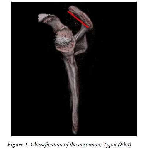
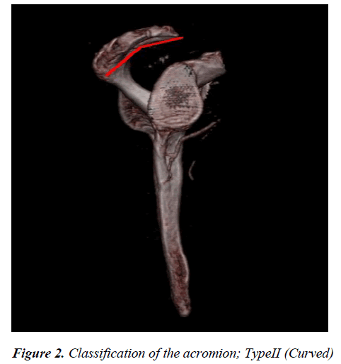
In our study, after morphological analysis we observed three diffent acromion type which has reported in previously conducted study. We found in 55 (37%) subjects with flat acromion which is known as Type I (Figure 1), in 71 (48.7%) subjects with curved acromion
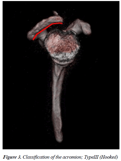
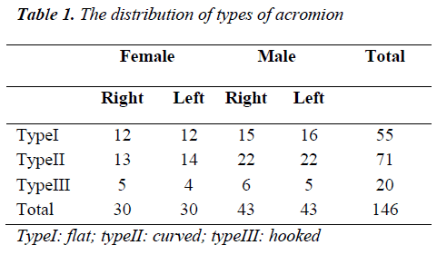
which is known Type II (Figure 2) and in 20 (13.7%) subjects with hooked acromion which is known as Type III (Figure 3). Morphological classification of the acromion according to gender and lateralization is given with all details in Table 1.
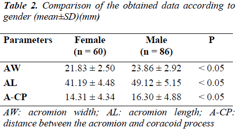
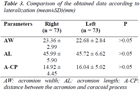
After morphological measurements, it is determined that AW is 21.83 ± 2.50 mm in woman and is 23.86 ± 2.92 mm in men. while AL is 41.19 ± 4.48 mm in woman and 49.12 ± 5.15 mm in men, the distance between A and CP is determined as 14.31 ± 4.34 mm in women and 16.30 ± 4.88 mm in men. The significantly difference were found between gender in all of the three parameter (p<0.05) (Table 2). The value of the parameters was recorded for both right and left side (Table 3) and no statistically significant difference was found between these values (p>0.05).
Discussion
Tendinitis which occurs as a result of compression of rotator cuff tendons under coracoacromial arc is one of the most common causes of shoulder pain. Supraspinatus tendon may be compressed by humerus head and coracoacromial arc during shoulder joint elevation. One of the important etiological factors for appearance of this syndrome is morphological modifications in the acromion [2]. Researches conducted reveals the relation between acromion morphology and subacromial compression syndrome and rotator cuff ruptures which causes shoulder pain [4,7-10]. The slope on the acromion end compresses on the supraspinatus tendon and the purpose of the surgery is to widen supraspinatus exit by obtaining a flat area [11].
First, Bigliani et al. [12], have classified acromion as type I (flat), type II (sloped to anterior) and type III (hook). Wang et al. [13] have classified acromion types in two groups under and above 50 years. Type I, type II and type III have been detected by 43.3.%, 45.2% and 11.5%, respectively in individuals younger than 50 whereas type I, type II and type III have been recorded as 32.8%, 43.7% and 23.5%, respectively in individuals older than 50. In the study conducted by Balke et al. [6] on 50 scapulae, they have detected type I acromion as 18%, type II acromion as 80% and type III acromion as 2%. Paraskevas et al. [14] have detected type I acromion as 26.1%, type II acromion as 55.6% and type III acromion as 18.1%. In our study, type I acromion was detected by 38%, type II acromion was detected by 49% and type III acromion was detected by 13%. Our findings complied with literature findings and type II acromion rate was the highest, type I and type III acromion rates followed these.
In the study of Crahım et al. [15], acromion morphology has been emphasized in the etiology of subacromial compression syndrome and detected acromion width as 3.68 ± 1.39 cm and acromion length was detected as 3.70 ± 2.66 cm. Collıpal et al. [7], have detected acromion width in their study as 25.12 ± 1.8 mm on the right side and 25.12 ± 1.8 mm on the left side. In the same study, acromion length was recorded as 69.12 ± 3.5 mm on the right side and 63.15 ± 7.1 mm on the left side. According to this study, acromion length between right and left side was found statistically significant. In our study, acromion width on the right side was 23.36 ± 2.99 mm on the right side and 22.68 ± 2.84 mm on the left side; acromion length was detected as 45.99 ± 5.90 mm on the right side and 45.72 ± 6.62 mm on the left side. Acromion width values obtained in our study comply with results of Collıpal et al. [7]. Other results of ours are different from the literature. The difference between our results on the right and left side is not statistically significant despite Collıpal et al. [7]. However, the difference between the genders is statistically significant in both parameters.
Rotator cuff adheres onto the humerus by passing from the narrow gap between the humerus head and coracoacromial arc. Any kind of mechanical factors narrowing the supraspinatus exit causes friction and abrasion on the tendon [3]. Coracoacromial distance is an important factor to understand etiology of shoulder pains. Wide gap decreases the risk of pain due to this region [1]. Narrower gap causes more risk for occurrence of rotator cuff ruptures [6]. In the study of Ünal et al. [16], coracoacromial distance was recorded as 21.4 mm in the narrowest place and as 33.3 mm in the widest place. Collıpal et al. [7] have reported this distance as 39.76 ± 5.2 mm on the right and 39.55 ± 5.4 mm on the left and they have mentioned that the difference between two sides are not statistically significant. Alpyörük Taşer et al. [1], have reported coracoacromial distance as 30.8 ± 4.5 mm in female patients and 35.5 ± 3.7 mm in male patients. The difference between the genders is statistically significant. In our study, the narrowest distance between acromion and coracoid process was detected as 14.31 ± 4.34 mm in females and 16.30 ± 4.88 mm in males. We believe that the cause for lower values detected in our study when compared with the values in the literature is due to the measurement from the narrowest site of coracoacromial distance. Furthermore, since our findings are close to those detected by Ünal et al. [16], this narrow distance may be a characterictics of Turkish community.
Because deltoid adherence site is completely preserved in acromioplasty surgery which creates basic philosophy of arthroscopic subacromial decompression, it is more advantageous than open techniques [11]. To know anatomic details and variations of the region is important for diagnosis and treatment. Furthermore, to recognize that important parameters of the region may change according to the race, gender and lateralization will significantly increase the surgical achievement. Therefore, we believe that data that we revealed would be useful for orthopedists in differential diagnosis and surgical interventions of shoulder pains. Furthermore, we think that our study is useful for creation of statistical and reference information of Turkish community.
References
- Alpyörük Taşer F, Başaloğlu H. Morphometrıc dımensıons of the scapula. Ege tıp dergisi 2003; 42:73-80.
- Akman Ş, Küçükkaya M. Subacromial impingement syndrome: Pathogenesis, clinical features, and examination methods. Acta Orthop Traumatol Turc 2003; 37: 27-34.
- Atalar AC, Demirhan M, Kocabey Y, Akalın Y. Arthroscopic subacromial decompression: one- to seven-year results. Acta Orthop Traumatol Turc 2001; 35: 377-381.
- Gill TJ, McIrvin E, Kocher MS, Homa K, Mair SD. Hawkins RJ The relative importance of acromial morphology and age with respect to rotator cuff pathology. J Shoulder Elbow Surg 2002; 11: 327-330.
- Kesmezacar H, Babacan M, Ergüner R, Öğüt T, Cansu E. The value of acromioplasty in the treatment of subacromial impingement syndrome. Acta Orthop Traumatol Turc 2003; 37: 35-41.
- Balke M, Schmidt C, Dedy N, Banerjee M, Bouillon B, Liem D. Correlation of acromial morphology with impingement syndrome and rotator cuff tears. Acta Orthop 2013; 84: 178-183.
- Collıpal E, Sılva H, Ortega l, Espınoza E, Martıınez C. The acromion and its different forms. Int Jmorphol 2010; 28: 1189-1192.
- Mansur DI, Khanal K, Haque MK, Sharma K. Morphometry of acromion process of human scapulae and its clinical importance amongst Nepalese population. Kathmandu Univ Med J 2012; 10: 33-36.
- Toivonen DA, Tuite MJ, Orwin JF. Acromial structure and tears of the rotator cuff. J Shoulder Elbow Surg 2012; 4: 376-383.
- Zuckerman JD, Kummer FJ, Cuomo F, Greller M. Interobserver reliability of acromial morphology classification: an anatomic study. J Shoulder Elbow Surg 1997; 6: 286-287.
- Karaman Ö, Karakuş Ö, Kaynak G, Çalışkan G, Saygı B. Artroskopik subakromiyal dekompresyon: 1-4 yıllık sonuçlar. Şişli Etfal Hastanesi Tıp Bülteni 2012; 46: 136-139.
- Bigliani LU, Morrison DS, April EW. The morphology of the acromion and its relationship to rotator cuff tears. Orthop. Trans 1986; 70: 228.
- Wang JC, Shapiro MS. Changes in acromial morphology with age. J Shoulder Elbow Surg 1997; 6: 55-59.
- Paraskevas G, Tzaveas A, Papaziogas B, Kitsoulis P, Natsis K, Spanidou S. Morphological parameters of the acromion. Folia Morphol (Warsz) 2008; 67: 255-260.
- Crahım LF, Nagato AC, Rocha CLJV, Sılva MAS, Bandeıra ACB, Ferreır ATA, Bezerra FS. Acromial morphometric analysis using imaging software. Int J. Morphol 2013; 31: 345-350.
- Ünal N, Özden H, Özçelik A, Akdoğan I, Günal I. Subakromial aralık, akromion kalınlığı ilişkisi. Morfoloji Dergisi 1997; 5: 10-12.
