- Biomedical Research (2015) Volume 26, Issue 4
The effect of prescription of different Dexamethasone doses on reproductive system
Abbas Arab Dolatabadi*, Saeid Rezaei ZarchiiDepartment of Biology, Payame Noor University, I.R. Iran
Accepted : May 10 2015
Abstract
Dexamethasone is a widely used glucocorticoid which has been prescribed increasingly in recent years. The effects of Dexamethasone on LH, FSH and testosterone levels were investigated in present study. In this experimental work, 40 adult male Wistar rats were assigned into four tenmembered groups. The rats in the control group received saline, while the rats in experimental group were subjected to intraperitoneal injection of dexamethasone in 0.4, 0.7, and 1 mg/kg doses daily for period of 10 days. On following day of last injection, the rats were anaesthetized and plasma was drawn from their heart, FSH, LH and testosterone levels were measured and the results were analyzed using SPSS software and Dunnett test. The hormonal test of LH, FSH and testosterone was made using Pars kit and the groups were compared. In this study, no significant change was observed in the targeted hormones up to 0.7mg/kg dose, but significant changes in the concentration of LH, FSH and testosterone were found in higher doses, i.e., 1 mg/kg dose in experimental group compared to the control group (P<0.05). The results of present study indicated that frequent injection of dexamethasone has come up with multiple changes in LH, FSH and testosterone and it has borne harmful effects on the reproductive system of male rats.
Keywords
Dexamethasone, testosterone, Follicle-stimulating Hormone, Luteinizing Hormone.
Introduction
Reproductive function in primates and rats are controlled by somatic and mental stresses [1]. Studies indicate that an increase in stress-induced glucocorticoids results in decrease in serum concentration of testosterone in males [2]. Glucocorticoids have a wide therapeutic range includes inflammatory, infectious and autoimmune diseases, adrenal gland failure, joint pain and lymphoid cancers. Due to the wide therapeutic range of these compounds, excessive prescription of which by physicians has been obvious in recent years [3]. Glucocorticoids bring about the inhibition of testosterone by influencing anterior pituitary and testes [4]. When the testes are not able to support spermatogenesis through the production of testosterone, a special message transfer path is activated that ultimately results in apoptosis [5]. Glucocorticoids are strong medicines that are used mainly for inflammatory disorders such as asthma, allergy, infection as well as autoimmune diseases such as rheumatoid arthritis, Glomerulonephritis, sclerosis and Systemic Lupus Erythematosus [6] . Dexamethasone is a synthetic glucocorticoid whose potent to suppress the immune system is 20-30 times greater than that of hydrocortisone [7]. Glucocorticoid is rapidly diffused in the circulatory system and engender the regulation associated with transcription of some genes while passing through cell membrane. Glucocorticoids silence the genes that have a major role in inflammation such as cytokines as well as inflammatory enzymes such as nitric oxide synthase [8]. Cytokines make inactive the inflammatory enzymes such as nitric oxide synthase [8]. Based on available potent evidences, cytokines can directly stimulate the development of ROS [9,10]. Glucocorticoids are the main hormones secreted from adrenal gland. These hormones act as very strong anti-inflammation and inhibitors of immune system. This property has made them as the most-prescribed drug throughout the world [11].
The plasma level of testosterone is provided by steroidmaking process of testes’ leydig cells. leydig cells synthesize glucocorticoid receptors. Therefore, the first target of glucocorticoids’ activity is testicle tissue [12]. Dexamethasone is a highly used glucocorticoid whose prescription is made increasingly [13]. This drug is bound to its receptors in cytoplasm by passing through cell membrane and then enters the cell nucleus by drug-receptor complex. Through binding to specific areas of DNA, this complex leads to stimulation of mRNA transcription and subsequently development of enzymes that are ultimately responsible for systematic effects of corticosteroids. Dexamethasone applies its anti-inflammatory effects by preventing the accumulation of inflammatory cells in the inflammation zone, inhibition of phagocytosis and diffusion of enzymes responsible for inflammation and inhibition of production and dissemination of chemical mediators relevant to inflammation [14]. Dexamethasone is highly used and the reports by Medical Science Universities in Iran indicates high and increasing prescription of dexamethasone in recent years [15]. Estradiol binds to estrogen receptor in cytoplasm and promotes the production of DNA, RNA, and other proteins in target tissue. Further, the level of GnRH in hypothalamus declines under the effect of estrogen and the release of LH and FSH decreases in pituitary, although, its major role is in reproductive tissues such as uterus, but it also plays a role in other tissues such as osseous, hepatic, vascular and cerebral. Estradiol is produced in trace amounts from Testosterone metabolism. Laydig cells in testicles secrete testosterone while triggered by LH from anterior pituitary. Likewise, secreted testosterone level has rather direct relationship with available LH hormone.
The released testosterone from testicles in response to LH has a mutual effect in inhibition of LH release from anterior pituitary [4]. Major part of this inhibition is raised from direct effect of testosterone in reduction of GnRH secretion from hypothalamus. This leads to corresponding decline in LH and FSH release from anterior pituitary and decrement in LH, lessens the testosterone release from testicles. Thus, over-secretion of testosterone acts on hypothalamus and anterior pituitary via a negative feedback that turns back testosterone level to normal status. In contrast, below normal level releasing of testosterone leads to secreting high level of GnRH from hypothalamus, and it subsequently enhances LH and FSH release from anterior pituitary as well as testosterone secretion from testicles [4]. Based on some reports, dexamethasone ampoule is the most prescribed drug in Iran in the past 5 years. Therefore, the effects of different concentrations of dexamethasone on sexual hormones have been explored in the present study. Thus, according to the fact that no exhaustive research has been carried out in regards with the effects of Dexamethasone on gonad spermatogenesis- pituitary axis as one of most complicated and active physiologic axes in living beings, cognition of factors affecting on which as well as prevention or intensification methods of these effects are of most importance.
Materials & Methods
Devices
Centrifuge device (eppendorf), electrochemiluminescence device. The biochemical tests were done using Elecsys 2010 electrochemiluminescence device made by Japanese HITACHI and German Roche companies.
Materials
Ketamine was used for anesthetizing rats. The laboratory kits for T, LH and FHS have been provided from Pars Azmoon Company which consists of reference laboratory verification.
Animals
This experimental study is made on 40 adult male rats with average weight of 230±20 g. For adaptation and consistency with the experiment’s conditions, the animals are kept in appropriate ambient temperature, light and heat of the nest for 2 weeks and then they are put under the treatment. The feeding of rats is performed by prepared standard materials including 20 percent protein, 50 percent starch, 10 percent cellulose, 15 percent fat and vitamins. The animals are allowed to access enough food and water throughout the experiment.
The animals are randomly assigned into four tenmembered groups including: the experimental groups 1, 2 and 3 that receive dexamethasone in 0.4, 0.7, and 1 mg/kg, respectively through intraperitoneal injection and group 4 that is considered as control group and will only receive placebo as 1 mL/day [16]. After the period, each rat was anesthetized by ketamine (50 mg/kg) and xylazine (4 mg/kg). In order to measure T, FSH and LH, 5 mL of serum will be drawn out of heart and studied using hormonal kits by Electrochemiluminescence.
Hormone analysis
The plasma was drawn out of the hearts of animals while 10 days are passed and the blood samples were centrifuged in 3000 RPM for 10 minutes and after separating serum, they were transferred to Electrochemiluminescence (Elecsys 2010) and LH (miu/ml), FSH (miu/ml) and testosterone (mg/ml) were measured.
Data analysis method
ANOVA test will be used for comparison of the level of sexual hormones in the control and experimental groups. The required software is SPSS and the charts will be drawn by Excel.
Results
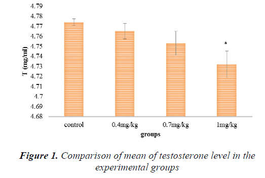
As it is shown in figure 1, the level of testosterone has been reduced in all experimental groups compared with the control group but this reduction is significant in the group receiving 1 mg/kg.
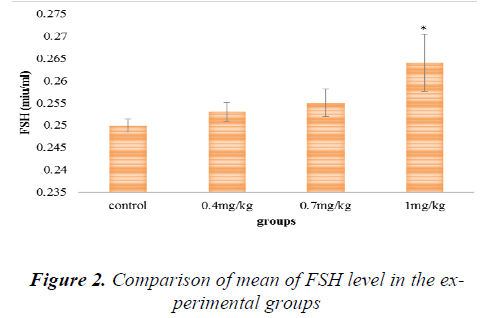
As it is shown in figure 2, the level of FSH has been increased in all experimental groups compared with the control group but this increase is significant in the group receiving 1mg/kg.
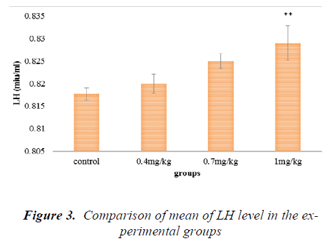
As it is shown in figure 3, the level of LH has been increased in all experimental groups compared with the control group but this increase is significant in the group receiving 1mg/kg.
Discussion
The present study indicated that interapertoneal injection of dexamethasone brings about the increase in FSH and LH and the reduction in testosterone and these changes are dose dependent and significant at the highest dose (1 mg/kg) (p<0.05). Pellegrini et al studied the effect of the stimulating drug ACTH and the inhibiting drug dexamethasone in synthesis steroid hormones in adrenal cortex on testes and epididymis of rats and showed that a clear histological damage has occurred in testes as well as epididymis [17]. Tsantarlotou et al explored the effects of dexamethasone in 3mg/kg concentration on acrosin activity in the sperm of Greek sheep during fall period and indicated that glucocorticoid receptors exist in epididymis and accessory glands [18] . The study by Silvia et al indicated that dexamethasone changes the glucocorticoid stimulant and reactive gene expression in the nuclei of epididymal epithelial cells and the epididymis is under the regulation of glucocorticoid [19]. The effect of dexamethasone on apoptotic proteins Fasl and Bax in sperm germ cells in the testes of mice was studied and the results showed that glucocorticoid compounds such as dexamethasone result in apoptosis and disruption in spermatogenesis by affecting pro-apoptotic proteins such as Fasl and Bax [19]. The researchers measured acrosin activity level in the samples taken from semen in 48 hours before the injections and during the use of dexamethasone in days 0.4 and 0.7. They also studied the level after the fulfillment of experiments every week up to 77 days following to the administration of dexamethasone. Also, testosterone sample was determined 24 hours before the injection and in the same injection days and in days 4, 7, 14 and 21 using immunoassay method. In this study it was found out that the most stimulation in the activity of acrosin in sperm had been emerged during days 7 to 21 following to the use of dexamethasone while the activity of acrosin before day 7 had been highly reduced. Also, the serum testosterone level showed a clear reduction. The researchers found that dexamethasone may have a direct effect on the synthesis and secretion of acrosin inhibitor in seminal fluid or epididymis as glucocorticoid receptors are present in epididymis and accessory glands and epididymis has a significant role in sperm maturation [20] .This is consistent with the results of the present study and testosterone level in the highest does of dexamethasone has been significantly reduced. Silvia et al studied glucocorticoid receptors in mice’s epididymis. In this study, treatment with dexamethasone on natural tissue was ex plored and a significant change occurred in the gene expression of stimulant and reactive gene expression in the nuclei of epididymal epithelial cells. This study indicate that epididymis is under the regulation of glucocorticoids [19]. Hashemitabar et al studied the effect of dexamethasone on Fas L apoptotic protein in testicular germ cells in the testes of 24 male mice and found that glucocorticoid compounds such as dexamethasone result in apoptosis and disruption in spermatogenesis process and significant reduction of spermatocytes in laboratory mice by influencing pro-apoptosis proteins such as Fas L [13]. In another study, Khorsandi et al studied the effect of dexamethasone on Bax protein expression as an apoptotic protein in germ cells of 35 male mice and found that glucocorticoid compounds such as dexamethasone can come up with apoptosis and disruption in spermatogenesis process affecting pro-apoptosis proteins such as Bax [20] . And also in this study, dexamethasone has led to damage to testes tissue and modification of testosterone in serum. In present study, administration of Dexamethasone promotes secretion of LH and FSH and attenuates Testosterone level in female mouse. Several studies showed that Corticosterone enhances the release of FSH in vitro and selective Corticosterone promotes mRNA subunit β-FSH in vitro. These studies support our results in vivo [21]. Prior studies on Dexamethasone influence on ovary function suggested that it inhibits the enhancement of FSH in Granulosa cells as well as Estrone secretion in vitro [22].
Conclusion
The results of the present study indicated that frequent injection of dexamethasone has resulted in multiple changes in LH, FSH and testosterone and it bears harmful effects on the reproductive system of male rats and this indicates that highly prescription and administration of this drug results in harmful effects on testicle tissue and alterations in sexual hormones. In summary, in present study, presence of Dexamethasone resulted in LH and FSH secretion in adult male mice in vivo. Enhancement of LH, FSH in treated mice with Dexamethasone, mediates the suppression of testicle function.
References
- Dong Q, Salva A, Sottas CM, Niu E, Holmes M,Hardy MP. Rapid glucocorticoid mediation of suppressed testosterone biosynthesis in male mice subjected to immobilization stress. Journal of andrology. 2004; 25(6):973-81.
- Hardy MP, GaoH-B, Dong Q, Ge R, Wang Q, Chai WR, et al. Stress hormone and male reproductive function. Cell and tissue research. 2005; 322(1):147- 53.
- Schmidt S, Rainer J, Ploner C, Presul E, Riml S, Kofler R. Glucocorticoid-induced apoptosis and glucocorticoid resistance: molecular mechanisms and clinical relevance. Cell Death & Differentiation. 2004; 11:S45-S55.
- Herrera‐Luna C, Budik S, Helmreich M, Walter I, Aurich C. Expression of 11β‐Hydroxysteroid Dehydrogenase Type 1 and Glucocorticoid Receptors in Reproductive Tissue of Male Horses at Different Stages of Sexual Maturity.Reproduction in Domestic Animals. 2013; 48(2):231-9.
- Koji T, Hishikawa Y, Ando H, Nakanishi Y, Kobayashi N. Expression of Fas and Fas ligand in normal and ischemia-reperfusion testes: involvement of the Fas system in the induction of germ cell apoptosis in the damaged mouse testis. Biology of reproduction.2001; 64(3):946-54.
- Rhen T, Cidlowski JA. Antiinflammatory action of glucocorticoids—new mechanisms for old drugs. New England Journal of Medicine. 2005; 353(16):1711-23.
- Mager DE, Moledina N, Jusko WJ. Relative immunosuppressive potency of therapeutic corticosteroids measured by whole blood lymphocyte proliferation. Journal of pharmaceutical sciences. 2003; 92(7):1521-5.
- Barnes P. Corticosteroid effects on cell signalling. European Respiratory Journal. 2006; 27(2):413-26.
- Corda S LC, Vicaut E, Duranteau J. Rapid reactive oxygen species production by mitochondria in endothelial cells exposed to tumor necrosis factor-a I smediated by ceramide. Am J Respir Cell MolBiol2001 Jun; 24(6):762-8.
- Ferro TJ, Hocking DC, Johnson A. Tumor necrosis factor-alpha alters pulmonary vasoreactivity via neutrophil- derived oxidants. American Journal of Physiology- Lung Cellular and Molecular Physiology. 1993; 265(5):L462-L71.
- Buckingham JC. Glucocorticoids: exemplars of multi‐tasking. British journal of pharmacology. 2006; 147(S1):S258-S68.
- Gametchu B, Watson CS. Correlation of membrane glucocorticoid receptor levels with glucocorticoid‐ induced apoptotic competence using mutant leukemic and lymphoma cells lines. Journal of cellular biochemistry. 2002;87(2):133-46.
- Hashemitabar M, Orazizadeh M, Khorsandi L. Effect of Dexamethasone on Fas Ligand Expression in Mouse Testicular Germ Cells. ZUMS Journal. 2008; 16(62):17-26.
- Katzung BG, Masters SB, Trevor AJ. Basic & clinical pharmacology. 2004.
- Soleimani F GK, Tehrani A. Increasing dexamethasone administration. Food Drug Commission 2003; 1: 3
- Du X, Liu J, Xin K, Liu G. Dexamethasone and sodium carboxymethylcellulose prevent postoperative intraperitoneal adhesions in rats. Brazilian Journal of Medical and Biological Research. 2015(AHEAD):000-.
- Pellegrini A, Ricciardi M. Histological and histochemicalobservations on the testis and epididymis of the albino rat treated with ACTH 1-39 and dexamethasone.International journal of tissue reactions.1982; 5(1):41-5.
- Tsantarliotou M, Taitzoglou I, Goulas P, Kokolis N. Dexamethasone reduces acrosin activity of ram spermatozoa. Andrologia. 2002; 34(3):188-93.
- Silva EJ, Queiróz DB, Honda L, Avellar MCW. Glucocorticoid receptor in the rat epididymis: expression, cellular distribution and regulation by steroid hormones. Molecular and cellular endocrinology. 2010; 325(1):64-77.
- Khorsandi LS HM, Orazizadeh M. The expression of BAX in mouse testicular germ cell apoptosis following Dexamethasone injection. Med Sci J Islamic Azad Univ 2008; 18 (3): 141-8.
- McAndrews JM, Ringstrom SJ, Dahl KD, Schwartz NB.Corticosterone in vivo increases pituitary follicle- stimulating hormone (FSH)-beta messenger ribonucleic acid content and serum FSH bioactivity selectively in female rats. Endocrinology.1994;134(1):158-63.
- Suzuki T, Miyamoto K, Hasegawa Y, Abe Y,Ui M, Ibuki Y, et al. Regulation of inhibin production by ratgranulosa cells. Molecular and cellular endocrinology.1987; 54(2):185-95.


