Research Article - Journal of Parasitic Diseases: Diagnosis and Therapy (2017) Volume 2, Issue 2
Occurrence of Gnathostoma spinigerum in tigers and leopards.
Singh KP*, Shrivastav AB, Gupta SK, Agrawal S, Singh K
School of Wildlife Forensic and Health, NDVSU Jabalpur, India
- *Corresponding Author:
- Dr. Keshav Pratap Singh
School of Wildlife Forensic and Health Jabalpur, India
Tel: 94253-59365
E-mail: kpsinghbaghel@yahoo.com
Accepted May 31, 2017
Citation: Singh KP, Shrivastav AB, Gupta SK, et al. Occurrence of Gnathostoma spinigerum in tigers and leopards. J Parasit Dis Diagn Ther. 2017;2(2):1-3
Abstract
The occurrence of Gnathostomiasis has been reported during post mortem examination of carcasses of 3 free-range tigers and two leopards from different Tiger Reserves of central India. Pathological lesions were characterized by bunches of nodules in the pyloric part of the stomach filled with light reddish colour worms with chronic gastritis. The parasites observed in the colony with 1:9 ratios of male and female worms measured 10-26 mm × 1.8-2.2 mm males and 25-35 mm × 1.8-2.3 mm females that were collected and kept in normal saline. The morphological observation of worms and eggs were envisaged for species identification. The recovered parasites from carcasses of leopards and tigers were identical to the morphological features of Gnathostoma spinigerum. The parasite have potential for zoonotic migration as the aquatic copepods act as their primary intermediated host and fishes harbour the encysted larval stage of the parasite may lead the occurrence of infection mostly in carnivores while accidentally infect the human beings. Such infection has already been reported in eastern Indian tribes resides in the close vicinity of the tiger reserves and national parks thus the migration of zoonotic disease takes place through common water resources of sylvatic and domestic cycle vice-versa.
Keywords
Biometry, Bengal tiger, Panther, Gnathostoma spinigerum, Gastric tumour, Zoonotic transmission, Parasitic disease, Tribes, Protected forests.
Introduction
The role of wild animals in transmission of parasitic zoonotic infection is well ascertained owing to the expansion of human rehabilitation in the adjoining areas of the protected forests through admixture of the feral and wild animals [1]. However, indiscriminate uses of forest wealth resulting into squeezing of wilderness in addition to frequent uses of contaminated water resources either by carnivores or tribal peoples may pick up the infection of Gnathostomiasis. The problem persists particularly in carnivores owing to their dependency on aquatic fauna mostly in cubs of big cats [2] while such parasitic diseases have potentials to overcome the agility and alertness in infected carnivore as the Gnathostomes are voracious blood feeder [3].
Gnathostomes are Spirurids nematodes having indirect life cycle and widely prevalent in domestic and wild carnivores. Owen [4] reported Gnathostoma spinigerum in gastric fundus of a young tiger at London Zoo. The prevalence of G. spinigerum recorded from Asia, Australia, South America, and North America [3]. The life-cycle of Gnathostomiasis begins with the eggs that hatches into encysted larvae in Cyclops and usually eaten by fresh water fishes or by several species of frogs and reptiles whereas the infective larvae may also sustain as paratenic hosts in variety of mammals i.e., mice, rats, and primates etc. Habit and habitat seeking to prey predator relationship between herbivores and carnivores, the adult parasite develop in the stomach of definitive host and mature in the stomach wall in about six months [5]. From India, it was reported earlier in Tigers [1-11] while zoonotic transmission through wild carnivores to humans and vice versa is still obscured. The present communication aimed with accounting morphological differences of the parasites encountered from tigers and leopards to observe whether they are same or having distinct features with G. spinigerum that have potentials for zoonotic transmission, thus useful for preventive measures to overcome the disease burden in different national parks and tiger reserves of Indian sub-continents.
Materials and Methods
Detailed post mortem examination of 3 free-ranging tigers and 2 leopards of either sex revealed occurrence of Gnathostomiasis in Tiger Reserves of central India brought to the laboratory of School of Wildlife Forensic and Health during the years 2012- 2016. As per the Wildlife Protection act 1972, the post-mortem examination of Schedule 1A wild animal viz. tiger leopard and lions shall be subjected to diseases diagnosis, forensics and organised post mortem before their funeral. The individual carcass was screened for collecting the biological materials for forensics as well as diseases diagnosis with pathological observations of the gastrointestinal tract; in all cases, the stomach observed with numerous nodules associated with lesions of gastritis. These nodules were ruptured and worms collected in the physiological saline prior to their further procession in hot 70% alcohol to stretch their body for measurements. Further, parasites of either sex were collected and kept in the laboratory made 5% lacto-phenol as clearing agent to increasing the transparency subjected to study the position of the genital and excretory organs. About 10 worms of either sex were kept in the incubator at 37°C for 72 h for morphometry and photography of the worms and eggs for the assessment of differences in the worms recovered separately from carcasses of leopards and tigers following the standard protocol [3].
Results and Discussion
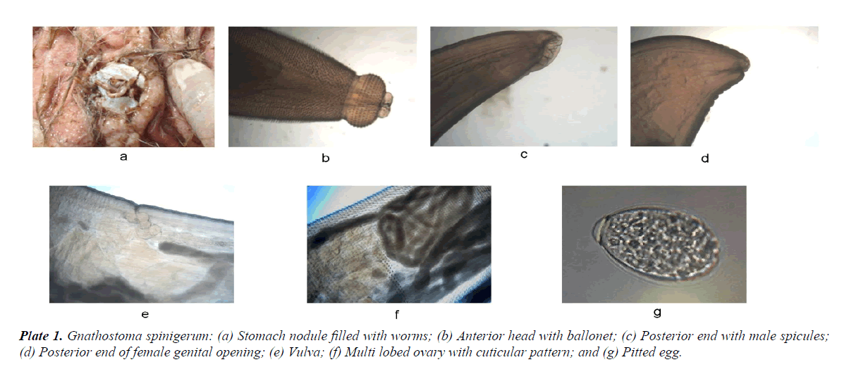
Macroscopically, the worms observed light-reddish in colour, cylindrical body, coiled in the bunch with a male and females located in the pyloric folds of the stomach formed nodules (Plate 1a). The anterior end of the worm of either sex posse’s head-bulb with four sub median cavity or ballonets (armed anterior head–bulb) with 8-10 transverse rows of simple hooks (Plate 1b). Observed cuticular spines/scale like notches throughout in the three forth part of the body with denticulate posterior edges (Plate 1f). The male worms measured rather shorter in length (10-26 mm × 1.8-2.2 mm) as compare to females (25-35 mm × 1.8-2.3 mm) with small spines and four pairs of large pedunculate papillae along with several smaller sessile (Plate 1c). The spicules (two in numbers) in male worms observed with unequal in length and laterally compressed projections located in the ventral region of the tail though female worms possess bilobed tail end (Plate 1d) with vulva opens 6-8 mm from the posterior end (Plate 1e). Furthermore, the female worms with multi lobed ovaries located in the mid region and huge number of eggs seen under low magnification (40X). These eggs were observed oval in shape and measured with the mean size of 65 μm × 52 μm having granulated homogenous embryonic masses (Table 1 and Plate 1) and spot at one end, like thin cap with colourless bright egg shell (Plate 1g).
| Observation | No Examined | Length (mm) | Width (mm) | Morphology | |||
|---|---|---|---|---|---|---|---|
| Head | Body | Tail | Genital organs | ||||
| Male | 10 | 10-26 (20 ± 5.8) | 1.8-2.2 (2 ± 0.4) | Head bulb armed with 4 ballonet and 8-10 rows of hooks | Large flat cuticle spines on anterior 2/3 part | Ventral caudal region with small spines and 4 pair’s pedunculate papillae. | Two unequal size spicules |
| (0.8-1.8 mm) | |||||||
| Female | 10 | 25-35 (29 ± 3.4) | 1.8-2.3 (1.9 ± 0.8) | Head bulb armed with 4 ballonet and 8-10 rows of hooks | Large flat cuticle spines with denticulate posterior edges | Ventral caudal region without spines and papillae. | Vulva opens in the 6-8 mm from the posterior end without flap |
| Eggs | 25 | 65-72 µm (67 ± 4) | 34-42 µm (36 ± 5) | Oval with thin cap called mucoid plug at one end, colourless bright shell with granulations | |||
Table 1: Morphology of eggs and worms of Gnathostoma spinigerum recovered from carcasses of Tiger and Leopards.
The results of biometry are more or less similar to as the morphological description provided for Gnathostoma spinigerum [3,4,11]. In the present study, the causes of death of the big cats including three tigers and two leopards were well documented with observation of gastric tumour followed by stomach perforation leading to some extent peritonitis, congestion in lungs and inflammation in the small intestine. More or less similar observation has also been encountered in the 19 years old tigress at Pench Tiger Reserve of Madhya Pradesh [2] with morphological details of the parasites and reported dehydration, anorexia and diarrhoea before death of the animals. However, in the present study emaciated carcasses were observed with chronic infection of Gnathostomiasis. The pathological legions followed by congestion and necrosis in the pyloric parts of the carcasses examined during post mortem [11] seems similar to the present study except they reported Ancylostoma braziliense whereas in all cases such parasites were not encountered. The possibility of transmission of the infection might be owing to eating of fishes and crabs during their young age as they learn hunting and recognizing the beasts of prey in close quarters in the presence of theirs mother [5]. Thus, once infection of G. spinigerum established inside the definitive host, there are limited therapeutic and preventive control measures and desperately the disease manifestation shows their ugly face as G. spinigerum to be a very pathogenic and fatal parasite of felines and canines forming gastric tumour and untimely death of the infected carnivores may takes place [8]. In such circumstances the Gnathostomiasis may lead to the infected animal up to the moribund stage owing to the less metabolic rate as the parasite reside in the stomach and affect the digestive mechanism [2]. Simultaneously, the zoonotic dissemination of the G. spinigerum infection [7] from a tribal lady of north east of Meghalaya with clinical symptoms of migratory cutaneous swelling and Eosinophilic Meningoencephalitis may be an alarm for those living in the close vicinity of forest adjoining areas and using common water holes for bathing and drinking of unclean water.
Hence, the common uses of water holes between sylvatic and domestic cycles may lead to migration of zoonotic infection and article may be helpful in surveillance of zoonotic diseases in protected and non-protected areas of the forest as well as their migration in human community.
Acknowledgement
The authors are thankful to Jitendra Agrawal, PCCF (Wildlife), Govt. of M. P. and Prof. (Dr.) P.D. Juyal, Vice-Chancellor, Nanaji Deshmukh Veterinary Science University, Jabalpur, India for their kind support in the present study.
References
- Shah HL. An integrated approach to study the zoonoses. J Vet Parasitol. 1987;1:7-12.
- Shrivastav AB, Singh KP, Bhat MA, et al. Occurrence of Gnathostoma in free range tigress. J Para Dis (Springer). 2011;35:75-6.
- Shrivastav AB. Wildlife health a new discipline: Essential for Tiger Conservation Programme. Intas Polivet. 2001;2:134-6.
- Levine ND. Nematode parasite of domestic animals and man. In: Burgess Publication (2nd edn.). W. Minneapolis, Minnesota, 1980.
- Prater SM. The Book of Indian animals. In: BNHS, Bombay, 1998.
- Arora BM, Prasad SA. Gnathostomiasis in a tiger (Panthera tigris). Indian J Vet Pathol. 1989;13:106-7.
- Barua P, Hazarika NK, Barua N, et al. Gnathostomiasis of the anterior chamber. Indian J Med Micro. 2007;25:276-8.
- Chandler AC. A contribution of the life- history of a Gnathostome. Parasitol. 1925;17:237-44.
- Owen R. Anatomical descriptions of two species of entozoan from the stomach of a tiger (Felis tigris linn) one of which forms a new genus of Nematoide, Gnathosotoma. Pro Zool Soc London. 1836;4:123-6.
- Soulsby EJL. Helminths, Arthropods and protozoa of domesticated animals. In: ELBS and Bailliere Tindall (7th edn.), London 1982.
- Thilakan NJ, Selvaraj J, Senthil Kumar S, et al. Concurrent infection of Gnathostoma spinigerum and Ancylostoma braziliense in tigress. J Vet Parasitol. 2007;21:191-2.
