Research Article - Biomedical Research (2018) Volume 29, Issue 2
RETRACTED: Expression of BKca in mesenteric artery smooth muscle cells in preeclamptic patients
Hua Zhong1#, Wei Liu1#, Yanjiao Li2, Jing Wen3, Yan Zhu2, Xiaoyu Hou2, Li Liu1 and Xiaodong Fu1*
1Department of Obstetrics, the First Affiliated Hospital of Southwest Medical University, Luzhou, Sichuan Province, PR China
2Perinatal Medicine, Southwest Medical University, Luzhou, Sichuan Province, PR China
3Laboratory of Cardiomyocytes, Southwest Medical University, Luzhou, Sichuan Province, PR China
#These authors contributed equally to this work
- *Corresponding Author:
- Xiaodong Fu
Department of Obstetrics
The First Affiliated Hospital of Southwest Medical University, PR China
Accepted date: October 31, 2017
Abstract
This study aimed to study the expression of BKca in mesenteric artery smooth muscle cells in patients with Preeclampsia (PE), and to explore the relationship between BKca electrophysiological activity and PE. Human mesenteric smooth muscle cells with good activity were obtained by acute enzyme method. The BKCa current was recorded by single channel patch clamp technique. Higher quality cells required for experiments were obtained, and the recorded channel currents were in accordance with the general properties of BKca. Under the cell-attached patch, when the voltage was clamped at +40 mv, +50 mv, +60 mv, the opening Probability (Po) of PE group was significantly lower than that of the normal pregnancy group, and the difference was statistically significant (n=20, p<0.05). Under the inside-out patch, the Po of PE group was significantly lower than that of the normal pregnancy group, and the difference was statistically significant (n=20, p<0.05). Under the inside-out patch, when the membrane potential was +40 mv, the bath by adding different concentrations of free Ca2+ (10-8 M, 10-7 M, 10-6 M and 10-5 M), the sensitivity of PE group to Ca2+ concentration was significantly lower than that of the normal pregnancy group (n=20, p<0.05). The activity of BKca in mesenteric artery smooth muscle cells of preeclamptic patients was weakened. Our findings suggested that the changes of BKca channel activity in mesenteric artery smooth muscle cells were related to the occurrence of PE.
Keywords
Large conductance calcium-activated potassium channel, Human mesenteric artery, Vascular smooth muscle cells, Preeclampsia
Introduction
Large conductance calcium- and voltage activated potassium channels are ubiquitously expressed and regulate a diverse array of physiological processes [1-3]. Disruption of BK channel function in human and animals is associated with a wide range of pathologies ranging from hypertension, autism, asthma, cancer, diabetes, obesity and other disorders of the vascular, nervous, endocrine and other systems [4,5]. BKca is the highest expression of potassium channels on vascular smooth muscle cells. When blood pressure rises, the high intracellular calcium can cause BKca activation, hyperpolarization of cell membrane, and hypervolemia smooth muscle relaxation. On the contrary, it can cause vascular smooth muscle contraction [6,7]. BKca plays an important role in regulating the relaxation and systolic function of vascular smooth muscle.
Preeclampsia (PE) is a human pregnancy-specific multi-system disease, with hypertension, proteinuria and edema as its major characteristics [8,9]. PE can cause endothelium damage and vascular spasm can lead to serious complications, such as acute renal failure, cerebral hemorrhage [10,11]. Thus, we supposed that BKca may be involved in the vascular spasm caused by PE.
Therefore, in the present study, we aimed to investigate the activity of BKca in mesenteric artery smooth muscle cells in patients with PE and to explore the correlation between BKca activity and PE, and to provide a theoretical basis for further understanding the etiology and pathogenesis of PE.
Materials and Methods
Patients and sample collection
The present study was approved by the Animal Ethics Committee of the First Affiliated Hospital of Southwest Medical University. Inform consent was obtained from every patient.
Specimens were collected from late pregnancy patients underwent cesarean section surgery in Luzhou Medical College Hospital from June 2013 to June 2014. Specimens were divided into the preeclampsia group (PE group, 20 cases) and normal pregnancy group (NLP group, 20 cases). PE group had an average blood pressure of 187.86 ± 8.13/101.43 ± 6.73 mmHg, and an average blood pressure of 114.34 ± 4.57/65.12 ± 5.42 mmHg was observed in NLP group. There was no significant difference in PE group and NLP group between gestational age and age.
Experimental reagents
Papain, bovine serum albumin, Dithiothreitol (DTT), XI collagenase, HEPES, K-aspartate and IbTX were all purchased from Sigma (Merck, Darmstadt, Germany).
Solution I: KCl 5.9 mM, NaCl 127 mM, HEPES 10 mM, glucose 12 mM, MgCl2 1.2 mM, CaCl2 2.4 mM.
Solution II: KCl 5.9 mM, NaCl 127 mM, HEPES 10 mM, glucose 12 mM, MgCl2 1.2 mM.
Single channel bath: KCl 40 mM, HEPES 10 mM, Kaspartate 100 mM, EGTA 1 mM.
Electrode liquid: KCl 100 mM, HEPES 10 mM, K-aspartate 40 mM, CaCl2 1mM.
Enzyme solution I: Papain 0.95 mg/ml, DTT 0.68 mg/ml, bovine serum albumin 2 mg/ml.
Enzyme II: Type XI collagenase 1.5 mg/ml
Acquisition of mesenteric artery smooth muscle cells
Tissues of the mesenteric A4~A5 segment with vascular was selected, and then cryopreservated with solution I. The small arteries with better elasticity and vessel diameter of 1 mm~2 mm were selected. After removing the surrounding fat and connective tissues, the small arteries were longitudinally opened, then the residual blood vessels and endometrial tissues were removed, and finally the small arteries were oblique cut into 2 mm × 2 mm tissue blocks. Combined with papain and type XI collagenase, the human mesenteric artery smooth muscle cells were isolated by using two-step acute enzyme method.
Single channel current recording
The mesenchymal smooth muscle cells isolated by acute enzymes were selected for cell-attached patch and inside-out patch experiments. The current signal was amplified by the patch clamp amplifier (CEZ-2200), 1KHz low-pass filter, the 12-bit A/D, D/A converter (Digidata-1200A Axon instrument USA), and then stored in the computer hard drive supported by the special software pClamp (Version 10.1, Axon Instruments, U.S.A). The sampling frequency was 10 KHz, sampling mode was Gap Free, and the sampling time was 30 s.
Statistical analysis
The experimental results were expressed as mean ± standard deviation (mean ± SD). SPSS 21.0 software was used to analyse the data. p<0.05 was considered as statistically significant.
Results
Artery smooth muscle cells isolated from human advanced mesenteric by two-step acute enzyme method
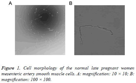
Using the two-step acute separation method, a large number of better conditions of mesenteric artery smooth muscle cells were obtained. Through the inverted phase contrast microscope, in the low magnification (10 × 10), each field of view can be seen in the number of cells 4~5 smooth muscle cells (Figure 1A). In the high magnification (100 × 100), the cell morphology was elongated shuttle type, such as earthworms, banana-like, occasional bifurcated Y (Figure 1B). Good smooth muscle cells are characterized by good adherence, cell membrane integrity, good refraction and strong sense of three-dimensional.
General characteristics of BKCa single channel in mesenteric artery smooth muscle cells of late pregnancy
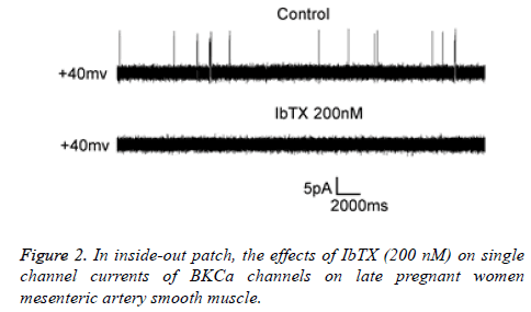
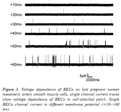
Scorpion Toxin (Iberiotoxin, IbTX) is a specific blocker for BKca, commonly used to identify BKca. In this experiment, under the inside-out patch, the bath by adding IbTX, the voltage clamp in the +40 mv, when IbTX was added to 200 nM, the single-channel current was almost completely blocked, proving that this current was BKca-mediated current (Figure 2). Under the attached patch, the activity of the channel exhibited a significant voltage dependency as the clamp current increased (Vm=0 to +60 mV) (Figure 3).
Comparisons of BKca open probability (Po) between PE group and NLP group
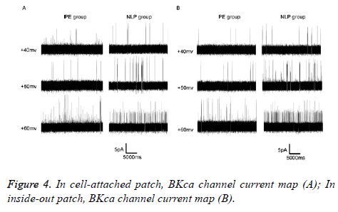
Under the cell-attached patch (Figure 4), when the voltages were clamped at +40 mv, + 50 mv, + 60 mv, the open probability (Po) of BKCa single channel current in NLP group was obviously larger than that of the PE group (Table 1).
| Clamp voltage | Po (PE group) | Po (NLP group) | T | P-value |
|---|---|---|---|---|
| +40 mv | 0.003 ± 0.000 | 0.010 ± 0.001 | 27.079 | 0 |
| +50 mv | 0.007 ± 0.000 | 0.032 ± 0.003 | 19.301 | 0 |
| +60 mv | 0.014 ± 0.001 | 0.051 ± 0.002 | 22.136 | 0 |
Table 1. The voltage dependence of BKCa channels from the three group in cell-attached patch (͞x ± s).
Under the inside-out patch, when the voltages were clamped at +40 mv, +50 mv, +60 mv, respectively, the open Probability (Po) of BKCa single channel current in NLP group was significantly larger than the open Probability (Po) of PE group, and the difference was statistically significant (p<0.05) (Table 2).
| Clamp voltage | Po (PE group) | Po (NLP group) | T | P-value |
|---|---|---|---|---|
| +40 mv | 0.004 ± 0.000 | 0.013 ± 0.001 | 54.356 | 0 |
| +50 mv | 0.009 ± 0.000 | 0.037 ± 0.002 | 93.266 | 0 |
| +60 mv | 0.016 ± 0.001 | 0.058 ± 0.002 | 78.692 | 0 |
Table 2. The voltage dependence of BKCa channels from the three group in inside-out patch (͞x ± s, n=20).
Comparison of the dependence of BKca on calcium ion concentration
In inside-out patch, the membrane potential was +40 mv and increased with the free Ca2+ concentration in the bath (10-7 M, 10-6 M, 10-5 M, 10-4 M), and at the same Ca2+ concentration, the open Probability (Po) of BKca single channel current in PE group was significantly lower than that of the NLP group, the difference was statistically significant (P <0.05) (Table 3).
| Calcium ion concentration | Po (PE group) | Po (NLP group) | T | P-value |
|---|---|---|---|---|
| 10-7 | 0.006 ± 0.000 | 0.025 ± 0.001 | 67.175 | 0 |
| 10-6 | 0.019 ± 0.001 | 0.043 ± 0.002 | 49.92 | 0 |
| 10-5 | 0.022 ± 0.001 | 0.074 ± 0.003 | 64.71 | 0 |
| 10-4 | 0.115 ± 0.009 | 0.253 ± 0.030 | 19.48 | 0 |
Table 3. The Ca2+ dependence of BKCa channels of PE group and NLP group in inside-out patch (͞x ± s, n=20).
Discussion
Almost all vascular smooth muscle cells have BKca expression [12,13]. Keep the smooth muscle cell membrane potential in resting potential, also can provide an endogenous compensation mechanism to buffer vasoconstriction, especially when the vascular tension increased [14,15]. At present, the vasodilator function of vascular smooth muscle cell BKca has been widely recognized, BKca exerts its protective role by enhancing the activity to combat excessive vasoconstriction [16,17]. The results of this study showed that the BKca channel of mesenteric artery smooth muscle cells in PE patients under both the two kinds of single-channel current recording mode, the open Probability (Po) was all lower than that of the normal pregnant women. The sensitivity of the mesenteric artery smooth muscle cell BKca channel to calcium concentration of the PE patients was also significantly lower than that of the normal pregnant women. In hypertensive disorder complicating pregnancy, NO is inhibited and BKca activity is reduced [18]. The role of BKca in vascular smooth muscle cells is inhibited by vasoconstrictor substances, thus leading to vasoconstriction [19]. It has been reported that the BKca current density of uterine artery smooth muscle cells in sheep was reduced, and the decrease of BKca activity was associated with down-regulation of β1 subunit [20]. A previous study used the whole-cell patch clamp technique to record the potassium channel current of placental vascular smooth muscle cells in hypertensive disorder complicating pregnancy, and found that the resting membrane potential increased and the potassium channel current decreased.
The preliminary study of this subject found that the BKca activity of the placental artery smooth muscle cells in hypertensive disorder complicating pregnancy decreased, β1 sub-unit expression decreased, and the electrophysiological changes of BKca were consistent with the expression of β1 sub-unit. At the same time, researchers also found that the BKca activity of the uterine artery smooth muscle cells in severe PE patients decreased. The results of this study are consistent with previous studies on BKca in hypertensive disorder complicating pregnancy. Therefore, in PE disease, the release of various placental factors in pregnant women, can promote the activation of inflammatory response and vascular endothelial injury, break down the ion exchange mechanism of the vascular smooth muscle cell membrane channel, present small vasospasm, and also lead to increased blood pressure [21]. These findings suggested that the change of BKca activity in mesenteric artery smooth muscle cells was involved in the development of hypertensive disorder complicating pregnancy. In the present study, the activity of BKca was attenuated during gestational hypertension. However, the current study of BKca in hypertensive disorder complicating pregnancy was still less, thus, it cannot be considered that the BKca activity in pregnancy-induced hypertension is simply a state of inhibition. In hypertensive disorder complicating pregnancy, whether BKca is inhibited or activated, whether the activity of BKca has a process from compensatory to decompensation, whether the changes in BKca activity lead to the occurrence of pregnancy-induced hypertension, or the occurrence of pregnancy-induced hypertension lead to BKca activity changes, still need further study.
Acknowledgements
The present study was financially supported by Sichuan Provincial Department of Science and Technology-Analysis of the expression of large conductance calcium activated potassium channels in uterine vascular smooth muscle cells in hypertensive disorder complicating pregnancy (2012SZ0178).
Disclosures
All the authors declare that there were not any types of competing interests in current manuscript.
References
- Contreras GF, Castillo K, Enrique N, Carrasquel-Ursulaez W, Castillo JP. A BK (Slo1) channel journey from molecule to physiology. Channels (Austin) 2013; 7: 442-458.
- Wu SN, Chern JH, Shen S, Chen HH, Hsu YT. Stimulatory actions of a novel thiourea derivative on large-conductance, calcium-activated potassium channels. J Cell Physiol 2017; 232: 3409-3421.
- Fezai M, Ahmed M, Hosseinzadeh Z, Lang F. Up-regulation of the large-conductance Ca2+-activated K+ channel by glycogen synthase kinase GSK3β. Cell Physiol Biochem 2016; 39: 1031-1039.
- Lu T, Jiang B, Wang XL, Lee HC. Coronary arterial BK channel dysfunction exacerbates ischemia/reperfusion-induced myocardial injury in diabetic mice. Appl Physiol Nutr Metab 2016; 41: 992-1001.
- Wang YJ, Chan MH, Chen L, Wu SN, Chen HH. Resveratrol attenuates cortical neuron activity: roles of large conductance calcium-activated potassium channels and voltage-gated sodium channels. J Biomed Sci 2016; 23: 47.
- Zhao HC, Wang F. Exercise training changes the gating properties of large-conductance Ca2+-activated K+ channels in rat thoracic aorta smooth muscle cells. J Biomech 2010; 43: 263-267.
- Borbouse L, Dick GM, Asano S, Bender SB, Dincer UD, Payne GA, Neeb ZP, Bratz IN, Sturek M, Tune JD. Impaired function of coronary BKCa channels in metabolic syndrome. Am J Physiol Heart Circ Physiol 2009; 297: 1629-1637.
- Karumanchi SA, Maynard SE, Stillman IE, Epstein FH, Sukhatme VP. Preeclampsia: a renal perspective. Kidney Int 2005; 67: 2101-2113.
- Chaiworapongsa T, Chaemsaithong P, Yeo L, Romero R. Pre-eclampsia part 1: current understanding of its pathophysiology. Nat Rev Nephrol 2014; 10: 466-480.
- Huppertz B. Placental origins of preeclampsia: challenging the current Hypothesis, Hypertension 2008; 51: 970-975.
- Carter EB, Conner SN, Cahill AG, Rampersad R, Macones GA, Tuuli MG. Impact of fetal growth on pregnancy outcomes in women with severe preeclampsia. Pregnancy Hypertens 2017; 8: 21-25.
- Lu T, Ye D, He T, Wang XL, Wang HL, Lee HC. Impaired Ca2+ dependent activation of large-conductance Ca2+-activated K+ channels in the coronary artery smooth muscle cells of Zucker diabetic fatty rats. Biophys 2008; 95: 5165-5177.
- Cheng J, Mao L, Wen J, Li PY, Wang N, Tan XQ, Zhang XD, Zeng XR, Xu L, Xia XM, Xia D, He K, Su S, Yao H, Yang Y. Different effects of hypertension and age on the function of large conductance calcium- and voltage-activated potassium channels in human mesentery artery smooth muscle cells. J Am Heart Assoc 2016; 5: 003913.
- Hu XQ, Xiao DL, Zhu RH, Zhang LB. Pregnancy upregulates large-conductance Ca2+-activated K+channel activity and attenuates myogenic tone in uterine arteries. Hypertension 2011; 58: 1132-1139.
- Brereton MF, Wareing M, Jones RL, Greenwood SL. Characterisation of K+ channels in human fetoplacental vascular smooth muscle cells. PLoS One 2013; 8: 57451.
- Ledoux J, Werner ME, Brayden JE, Nelson MT. Calcium-activated potassium channels and the regulation of vascular tone. Physiology 2006; 21: 69-78.
- Zhang S, Luo N, Li S. Inhibition of BKCa channel currents in vascular smooth muscle cells contributes to HBOC-induced vasoconstriction. Artif Cells Nanomed Biotechnol 2016; 44:178-181.
- Napolitano M. Expression and relationship between endothelin-1 messenger ribonucleic acid (mRNA) and inducible/endothelial nitric oxide synthase mRNA isoforms from normal and preeclamptic placentas. J Clin Endocrinol Metab 2000; 85: 2318-2323.
- Yang Y, Li PY, Cheng J, Mao L, Wen J, Tan XQ, Liu ZF, Zeng XR. Function of BKCa channels is reduced in human vascular smooth muscle cells from Han Chinese patients with hypertension. Hypertension 2013; 61: 519-525.
- Sholook MM, Gilbert JS, Sedeek MH, Hester RL, Granger JP. Systemic hemodynamic and regional blood flow changes in response to chronic reductions in uterine perfusion pressure in pregnant rats. Am J Physiol Heart Circ Physiol 2007; 293: 2080-2084.
- Goto K, Kansui Y, Oniki H, Kitazono T. Upregulation of endothelium-derive hyperpolarizing factor compensates for the loss of nitric oxide arteries of Dahl salt-sensitive hypertensive rats. Hypertens Res 2012; 35: 849-854.


