Case Report - Otolaryngology Online Journal (2016) Volume 6, Issue 3
Ectopic Supernumery Intranasal Tooth
Shivakumar Thiagarajan*, Sambandan AP and Ranjith Gopalakrishnan
Mahatma Gandhi Medical College & Research Institute, Puducherry, India
- *Corresponding Author:
- Shivakumar Thiagarajan
Former Assistant Professor, Mahatma Gandhi Medical College & Research Institute, Puducherry, India
E-mail: drshiva78in@gmail.com
Received date: April 14, 2016; Accepted date: May 09, 2016; Published date: May 12, 2016
Introduction
The ectopic eruption of teeth into the intranasal cavity is a rare clinical entity. Commonly seen in palate and maxillary sinus, mandibular condyle, coronoid process and even in the orbit in the maxillofacial region [1]. The presence of teeth has been reported in the ovaries, testes, anterior mediastinum and the presacral region as well. Ectopic teeth can be supernumery, deciduous or permanent. With the cosmetic problems of the external approach, an endoscopic approach has become essential in the removal of these ectopic intranasal teeth.
Case report
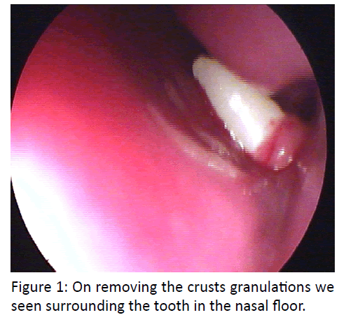
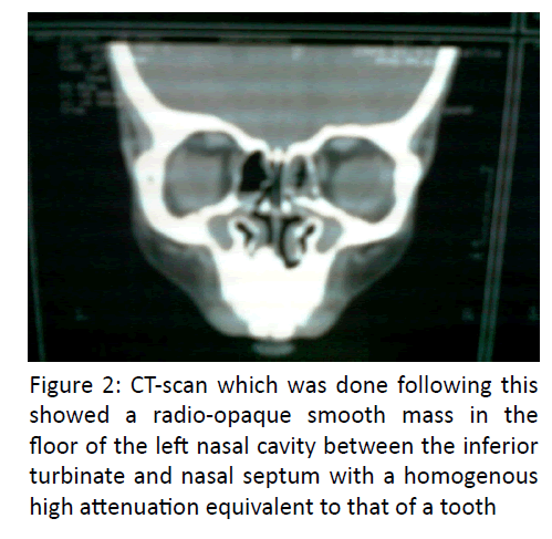
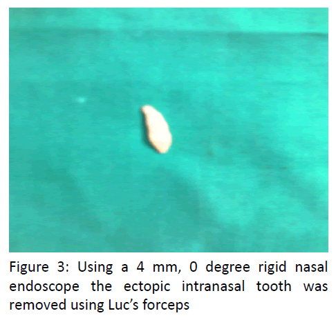
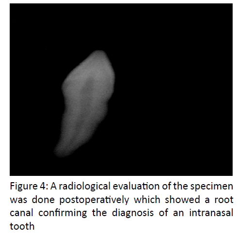
A 14 yrs old boy presented to the ENT OPD with nasal obstruction on the left side with occasional blood stained discharge for one month duration. He had no other nasal symptoms. There was however a history of fall with trauma to the upper (lateral) incisors at the age of 7 yrs. The patient’s general medical history was otherwise unremarkable. On anterior rhinoscopy a white mass with overlying crusts was seen on the nasal floor in the left side, on probing it was hard in consistency and immobile. Oral cavity examinations revealed a fractured upper lateral incisor on the left side, the remaining dentition were normal in appearance and number. Rest of the ENT examination was unremarkable. Subsequent to this a diagnostic nasal endoscopy was done which revealed a conical white projection tapering to a point superiorly from the floor of the left nasal cavity with crusts surrounding. On removing the crusts granulations we seen surrounding the tooth in the nasal floor (Figure 1). An orthopantamogram was taken which revealed that the patient had normal dentition. A CT-scan which was done following this showed a radio-opaque smooth mass in the floor of the left nasal cavity between the inferior turbinate and nasal septum with a homogenous high attenuation equivalent to that of a tooth (Figure 2). His routine blood and urine examination were within normal limits. He later underwent an endoscopic removal of the intranasal tooth under general anesthesia. Using a 4 mm, 0 degree rigid nasal endoscope the ectopic intranasal tooth was removed using luc’s forceps (Figure 3). The granulation and the remnant nasal mucosa surrounding the tooth were also removed and the base was cauterized. There was minimal bleeding during the entire procedure and the left nasal cavity was packed with medicated ribbon gauze, which was removed after 24 hrs and later the patient was discharged with medication. The patient is currently on follow-up and in good health. A radiological evaluation of the specimen was done postoperatively which showed a root canal confirming the diagnosis of an intranasal tooth (Figure 4).
Discussion
The incidence of supernumery teeth is between 0.1-1% of the general population. The most common location being the upper incisors, known as mesideons. The extra tooth has an atypical crown in vertical, horizontal or inverted position. The etiology of this ectopic intranasal tooth is not clear. Although the cause of ectopic growth is not well understood it has been attributed to obstruction at the time of tooth eruption secondary to crowded dentition, deciduous teeth or exceptionally dense bone [2].Other causes attributed are developmental disturbances such as cleft palate, rhinogenic or odontogenic infection and displacement as a result of trauma or cyst [2]. Multiple supernumery teeth are rare in individuals with no other associated diseases or syndrome [3]. Males are affected approximately twice as frequently as females [4,5]. Heredity may play a role, as supernumeraries are common in relatives of the affected children.
The diagnosis of intranasal is made on clinical and radiological findings. Clinically the patient may be asymptomatic or may present with nasal obstruction, epistaxsis, headache/facial pain, foul smelling nasal discharge, external nasal deformities, nasolacrimal duct obstruction [6,7]. On examination a white mass in nasal cavity is seen surrounded by granulation tissue and debris. Complication of intranasal tooth includes rhinitis caseosa, septal perforation or oroantral fistula [8]. Radiologically the nasal tooth appears as radio-opaque lesion with the same attenuation as that of oral teeth, as in our case. With bone window setting, the central radiolucency which is correlated with pulp cavity may have a spot or a slit depending on the orientation of the teeth. The soft tissue surrounding the radio-opaque lesion is consistent with granulation tissue found on clinical and pathological examination [9,10].
The differential diagnosis of intranasal tooth include, radio-opaque foreign body, rhinolith, inflammatory lesion with calcification (Syphilis, Tuberculosis or Fungal Infection), calcified polyp, osteoma, enchondroma, dermoid and malignant tumors like osteosarcoma and chondrosarcoma. However, the CT finding of the tooth equivalent attenuation and the centrally located cavity is confirmatory of the diagnosis of intranasal tooth [11].
Removal of the intranasal tooth is generally advised to alleviate symptom and to prevent complication. Endoscopic removal is advantageous in that it has good illumination, visualization and precise removal and is more convenient and safer than the traditional open methods, with reduced hospital stay and cosmetically satisfactory results. Asymptomatic tooth should also be removed, if not at least close radiographic follow-up is advised [11,12].
Conclusion
Intranasal tooth results from ectopic eruption of supernumery tooth and may cause a variety of symptoms and complications. An intranasal tooth is a rare clinical entity and the otolaryngologist should be aware of this while dealing with any nasal mass.
References
- Yeung KH and Lee KH (1996)Intranasal tooth in a patient with a cleft lip and alveolus. Cleft Palate Craniofacial. Journal33:157-159.
- Lee GS, Lee GY, Hong SL and Shin JK(1998)Supernumerary tooth in nasal cavity: report of 1 case. Korean Journal of Otolaryngol41:949-951.
- Lee FP (2001)Endoscopic extraction of an intranasal tooth: a review of 13 cases. Laryngoscope111:1027-1031.
- Carver DD, Peterson S and Owens T (1990)Intranasal teeth: a case report. Oral Surg. Oral Med. Oral Pathol70:804-805.
- Nastri AL and Smith AC (1996)The nasal tooth. Case report. Australian Dental Journal41:176-177.
- Medeiros AS,Gomide MR, Costa B, Carrara CF and das Neves LT (2000)Prevalence of intranasal ectopic teeth in children with complete unilateral and bilateral cleft lip and palate. Cleft Palate Craniofacial Journal37:271-273.
- Alexandrakis G, Hubbell RN and Aitken PA (2000)Nasolacrimal duct obstruction secondary to ectopic teeth.Ophthalmol107:189 -192.
- Moreano EH, Zich DK, Goree JC (1998)Nasal tooth.American Journal Otolaryngology 19:124 -126.
- Wurtele P and Dufour G (1994)Radiology case of the month: a tooth in the nose.J Otolaryngol 23:67-68.
- Albert Chen, Jon-Kway Huang, Sho-Jen Cheng (2002) Nasal teeth:report of three cases. American Journal of Neuroradiology 23:671-673.
- DaeHyungkim(2003) Endoscopic removal of an intranasal ectopic tooth. International Journal of Paediatric otolaryngology67:79-81
- Lt Col B Choudhry, Col AK Das (2008) Supernumery tooth in the nasal cavity.Medical Journal Of Armed Forces Of India 64:173-174.


