Research Article - Journal of Neuroinformatics and Neuroimaging (2017) Volume 2, Issue 2
Chronic low back pain (CLBP) is a disease; MRI signal change (Modic) in vertebrae is an active process by isotope bone scan.
Viroj Wiwanitkit*
Joseph Ayobabalola University, Akure Ilesha Rd, Ilara-Mokin, Nigeria
- *Corresponding Author:
- Viroj Wiwanitkit
Joseph Ayobabalola University
Nigeria
Tel: +234 807 761 1654
E-mail: wviroj@yahoo.com
Accepted date: 23 July, 2017
Citation: Alnaji A. Chronic low back pain (CLBP) is a disease; MRI signal change (Modic) in vertebrae is an active process by isotope bone scan. Neuroinform Neuroimaging. 2017;2(2):9-14
Abstract
Introduction: The pearl imaging study now days in evaluation of simple or complicated CLBP is the MRI of Lumbosacral spines. If we forget about prolapsed and dry discs as findings, here we confined our attention on a finding of a MRI signal change (Modic) +/- a dry disc or distorted disc with or without epiphyseal erosion.
Methods: 30 patients, 15 males and 15 females of 25-55 years old with chronic low back pain have MRI signal change and disc and epiphyseal distortion. Those patients are in a group considered as spondylitis according to the clinical basis.
Results: Showed different isotope accumulation rates but all considered positive for the accumulation. The magnitude of activity is linear with the degree of signal change or the structural change in MRI.
Conclusion: This could bring a hint on the biological bases of this signal change of MRI and hence the causation of CLBP in a group clinically diagnosed as bacterial spondylitis.
Keywords
MRI, Low back pain, Isotope bone scan, Modic, Spondylosis, Spondylitis, Brucella, Salmonella, Intracellular bacteria, Chronic infection, PCR, Micro-array, Comparative pathology, Failed back surgery.
Introduction
The signal or so simply the change of gray shade in MRI scan means a change in examined tissue characteristics, that’s to say a change in histological patterns, so it is a diseased tissue because any alteration in biological systems to right or towards the left is interpret in medicine as a disease regard less of the magnitude of this disease and causation. We are in a deadly need to discover this cause!
An area of the lumber vertebra happens to show this signal change with different sizes and patrons, some refer to it as a MODIC sign with many explanations to it. Below, the more agreed or the standard classification for vertebral body end plate MRI signal change, first described in 1988 [1,2].
Recently Modic type I has received renewed attention due to the possibility of it representing low grade indolent infection and is thus discussed separately [3].
Modic type I
• T1: Low signal
• T2: High signal represents bone marrow oedema and inflammation
• T1+C: Enhancement.
Modic type II
• T1: High signal
• T2: Iso to high signal represents normal red haemopoietic bone marrow conversion into yellow fatty marrow as a result of marrow ischaemia.
Modic type III
• T1: Low signal
• T2: Low signal represents subchondral bony sclerosis.
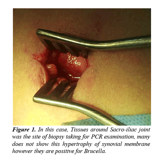
My work on the biological bases of the spondylosis (the structural alterations of any mode that affect all aspects of the vertebral column in spite of age or occupation or the abuse, some refer to as Osteoarthrosis of spine). In general, the last fifteen years showed that these structural changes are not due to progress in age because I have or more precise you find significant spondylosis in so young people even below nine years. Also not related to occupation or misuse or hard work also because you find it (spondylosis) in high percentage in people with so quite life. Spondylosis is a pathological finding found also in a wide spectrum of metabolically normal or apparently healthy people. What is left as a logic explanation is either tumor or a brand of infection. Radiological and clinically run with the operative and histopathological studies rule out the tumor causation. So what is left is the infection which I insist on. Based on strict clinical bases for diagnosis and the accordingly trial treatment with the excellent positive results, this career showed in thousands of patients with chronic low back ache is due to a chronic Brucellosis. The nearly to be a 100% positive rate of successful trial treatment for being a chronic Brucellosis is a fact. When the PCR tissue biopsy was entered into the service of diagnosis in the last three years showed 25% positive when the biopsy was taken from the sacroiliac joint area and the more than 60% positive when I changed to the periscapular muscles biopsy. The negative ones either are false negative or other intracellular bacteria like Salmonella and many others with the clinical improvements are due to cross antibiotic responses however this does not explain the whole issue with a comfortable satisfaction (Figure 1).
Method and Participants
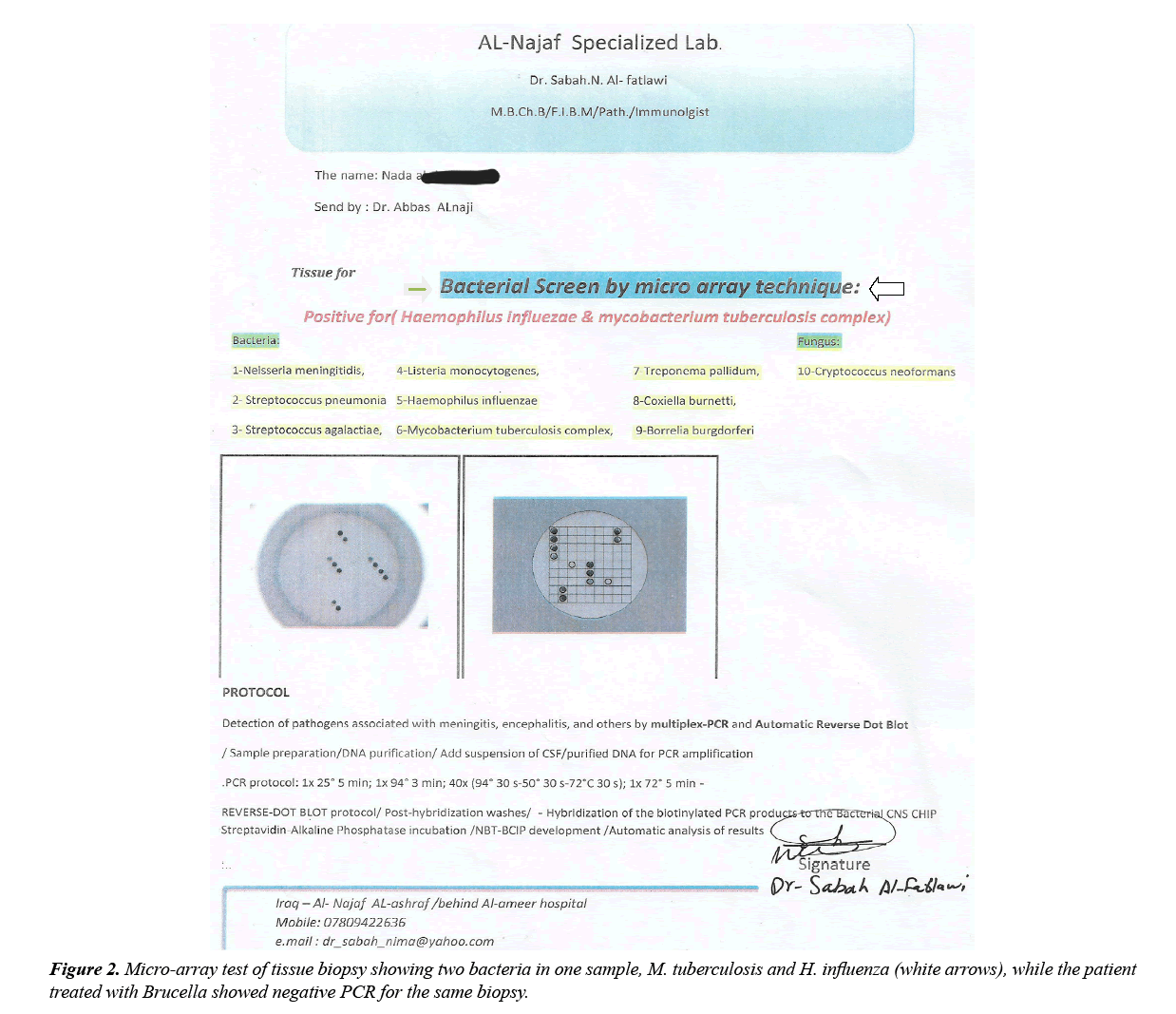
As mentioned earlier that this research was adopted some fifteen years ago and involve all patients (both genders and age groups starting from eight years to above) seen by me in public and private clinics in Iraq and some other countries where I worked as spine and a neurosurgeon. Full exclusive clinical and lab tests were done to exclude what is mentioned in standard textbooks as a cause for these spondylotic changes apart from PCR where it used in the last three years (Figure 2).
Findings and Results
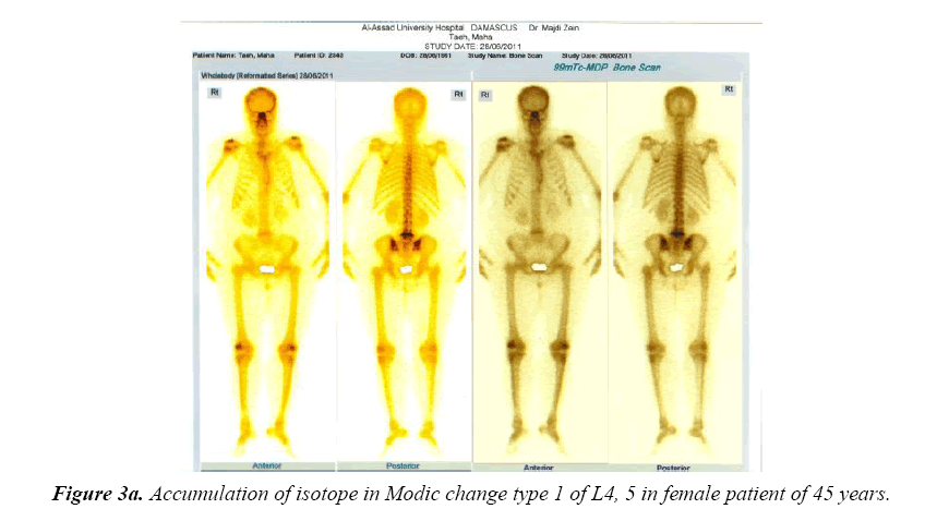
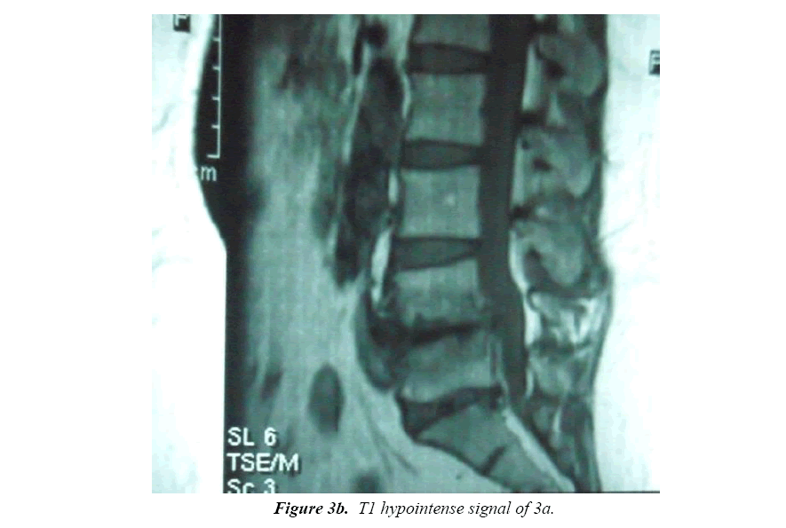
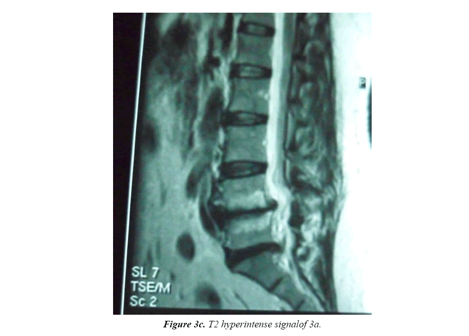
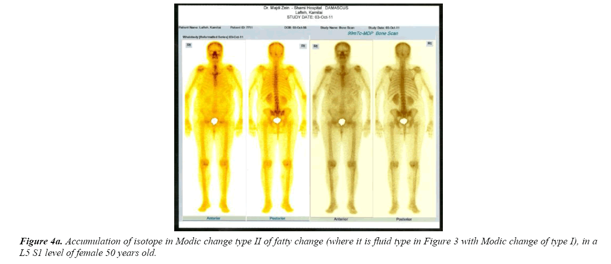
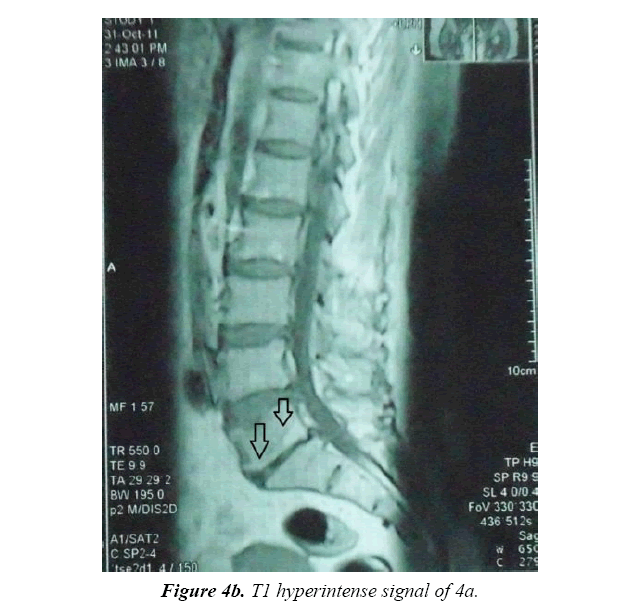
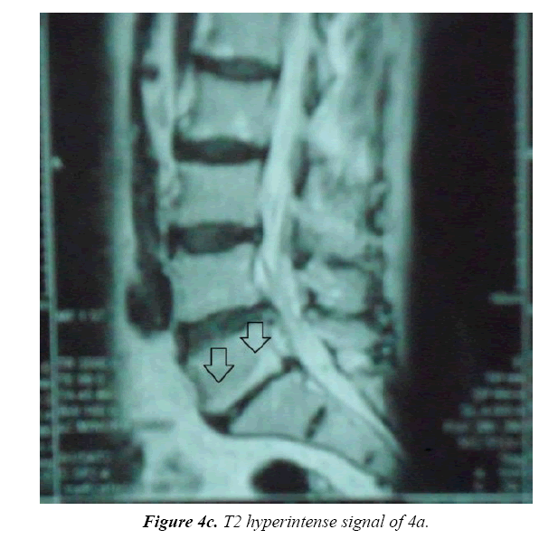
The spondylotic pictures in its various findings and ranges have been met over this long period with its related clinical pictures. Figures 3 and 4 showing the active tissue uptake of the isotope material and the correlated MRI images to show two various conditions but both took the radioactive material. Unfortunately we have no other modality of the changes which is the same in T1 and T2 as hypointense signal and refers to a sclerotic bone changes histologically.
Trial treatment based on clinical analysis was so excellent with regimens for chronic Brucellosis in the vast majority of the patients. Chronic Brucellosis was adopted due to the wide systemic affections the back pain in part or full spectrum is one of these findings but the major in those patients. Here, again I say unfortunately we lack the long term follow up of these patients, those showing their MRI and isotope studies here and the others because our patients disconnect their links to their doctors when become good and we hear about them indirectly from the patients sent by them for similar conditions this does not reflect the solid requirement or our needs.
Patients with the isotope scan (15 males and 15 females) had remarkable and progressive amelioration of their symptoms and signs on anti-Brucella without any type of analgesia, muscle relaxant, sedation or steroids, the symptoms and signs were made them candidate for surgery abolished with time. They did not underwent a tissue examination with PCR for Brucella because they did it before the administration of PCR in our service slightly more than three years from date of this article writing, they were obliged to travel to Syria where isotope scan was available but not in Iraq due to UN blockade, again unfortunately patients stop going to Syria those last three years to take the isotope scan due to recent ongoing war there. PCR tissue biopsy were 25% positive for Brucella from sacroiliac joint soft tissue and around 60% positive when the biopsy taken from pei-scapular muscles. Recently Micro-array biological molecular studies are now done with eight intracellular bacteria screen to detect the negative results with PCR for Brucella and to check whether if there is more than one invader like in this case (Figures 2, 3A-3C and 4A-4C).
Discussion
The huge number of patients over the long period with high positive outcome of treatment as Brucellar spondylosis (spondylitis) based on clinical analysis and trial treatment encouraged me to expand the vision to cover any chronic back aches under the tent of such findings. As for the spectrum of repeated or recurrent low backache is so wide ranging from simple or mild without clinical (physical) and radiological findings and even of very short duration may elicited by severe maneuver to a resting annoying pain do not responds to a seminarcotic like "Tramadol" intramuscular injections twice per day [4]. When both of such a fore mentioned severities respond with a very satisfaction when anti-Brucella is given it means nothing but the cause is this repeated low back pain is a complication to a chronic systemic low grade of sub-clinical brucellosis. The other features of chronic brucellosis when we look for and find them are a good support for this attitude especially when these other features which are also complications ameliorated within the course of treatment. As the cases with positive response increase with their wide range of features made this vision more nearer to the fact of low back pain is a complication to a chronic bacterial infections Brucella may be the most of being as an offending factor but it is under-estimated all over the world even if some workers isolated other pathogens. By this our understanding to the acute back ache should be re-directed to it is a pre-existing silent ongoing infection when some exertion practiced will elicit the pain just like when somebody eats a soft bread or drinks some cold water makes his tooth aches (comparative pathology to the same author) or sometimes it reaches a threshold to be aching without any precipitating factors. As matters go on untreated or managed with analgesia, rest, physiotherapy or even surgery in cases of sciatica due to prolapsed disc this sciatica even in presence of the prolapsed disc is faulty diagnosed when we omit the sciatic neuritis and surgeries are done with either right away no resolution or some honey moon intervals of relative comfort this reflects the inflammatory origin to the events in the neural canal skeleton, this principle includes the spinal canal stenosis where we find many laminectomies and foraminotomies do not solve the conditions whereas very good results obtained when anti-Brucella is given that the patient do not come to the second session of the treatment which is the surgery to correct the structural abbreviations done by the long standing bacterial effects because the reputation of back surgery all over the world is not without many drawbacks either instant or remote (could be the scientific explanation or the biological bases for failed back surgery) which this could be the sound analysis for it. From that it is the infection what spoils the structure of the spine whatever it is including the signal change (MODIC or others). We need to practice the biopsy taking from these signals changes not for histological or biochemical but for screen tests for the intracellular bacteria which are around fifteen in number other than Brucella.
So the three stages of these MRI signal changes (Modic I, II and III) represent the natural history of the ongoing bacterial vertebral end plate infection from the inflammatory edema in Modic I to the end process of tissue destruction and being replaced by fibrous (sclerotic) tissue in Modic III. It had been mentioned that Modic I is a bacterial infection in nature [5] but it seems that they expect that these isolated bacteria by cultures are due to the structural damage for some reason (you find here we returned to the chicken and the egg which from which!!) this damaged disc or end palate will encourages the bacteria they isolated in some patients and specifically in Modic I only [6- 9]. Here I stress again on the first stage of my work with trial treatment with anti-Brucella based on clinical bases was to high percentage positive including patients with Modic and others without Modic changes. As those with Modic responds very well to trial treatment with anti-Brucella is is so wealthy to go on the second stage of the study which is MRI re-imaging the Modic I patients to see the intensity behavior and the next stage is to take direct biopsy from Modic area and sent to Micro array Molecular screen test for a fifteen possible intracellular bacteria could be the cause behind the vertebral complex [9] destruction rather to any other traditional causes as the pure mechanical or senile.
Conclusion
Many origins had been given to be a cause for the spondylosis that causing the chronic low back pain, some convinced to be unknown. According to the above it is included in a process of chronic active intracellular bacterial vertebral infections.
Recommendation
Interested workers are welcome to share knowledge and further maneuvers toward such target.
Acknowledgement
This work is dedicated to whom in touch with the unknown, scientists and genius researchers.
References
- Modic MT, Steinberg PM, Ross JS, et al. Degenerative disk disease: assessment of changes in vertebral body marrow with MR imaging. Radiology. 1988;166(1):193-9.
- Rahme R, Moussa R. The modic vertebral endplate and marrow changes: pathologic significance and relation to low back pain and segmental instability of the lumbar spine. American Journal of Neuroradiology. 2008;29(5):838-42.
- Hacking C, Radswiki. Modic type endplate changes. Radiopaedia.
- https://www.drugs.com/tramadol.html.
- Albert HB, Sorensen JS, Christensen BS, et al. Antibiotic treatment in patients with chronic low back pain and vertebral bone edema (Modic type 1 changes): a double-blind randomized clinical controlled trial of efficacy. Eur Spine J. 2013;22(4):697.
- Urquhart DM, Zheng Y, Cheng AC. et al. Could low grade bacterial infection contribute to low back pain? A systematic review. BMC Medicine. 2015;13(1):13.
- Manniche C, Jordan A. 10 years of research: from ignoring Modic changes to considerations regarding treatment and prevention of low-grade disc infections. Future Sci OA. 2016;2(2).
- Hamlyn P, Albert H. MAST - Modic Antibiotic Spinal Therapy. Spine Surgery London. 2013.
- Alnaji A. LOW BACK PAIN IS A DISEASE: Vertebral Complex Structure. 2nd International Conference on Spine and Spinal Disorders, At Rome, Italy. 2017.
