Case Report - Journal of Advanced Surgical Research (2017) Volume 1, Issue 1
Acute Thromboembolism in Spleen Due to Clot in Left Atrial Appendages in Rheumatic Mitral Stenosis
Jayesh V Trivedi MD*
Head of Department and Professor of Medicine, Adani Institute of Medical Science, Bhuj, Gujarat, India
- Corresponding Author:
- Jayesh V Trivedi
Adani Institute of Medical Science Bhuj Gujarat
India
Tel: 098240 76530
E-mail: drjvtrivedi@rediffmail.com
Accepted Date: February 22, 2017
Citation: Jayesh V Trivedi MD. Acute Thromboembolism in Spleen Due to Clot in Left Atrial Appendages in Rheumatic Mitral Stenosis. Adv Surg Res. 2017;1(1):13-14
Abstract
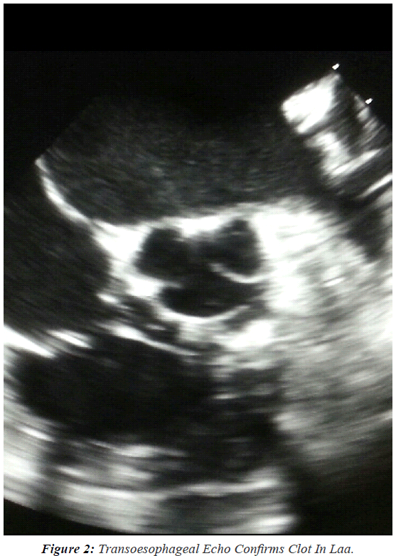
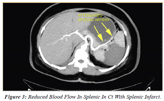
A 44-year-old woman was referred to our medical out door for control and management of acute abdominal pain in left hypochondrium in a patient who had Adenomyosis diagnosed by a Gynacologist. On examination patient was in sinus rhythm with auscultatory findings of Rheumatic Mitral Stenosis. She had soft rumbling mid diastolic murmur with presystolic accentuation and loud S1.Opening snap and loud pulmonary component of S2 was heard. Her Total WBC count was 15000 except this all her blood investigations were normal. She was complaining about left hypochondrium pain. Mild Splenomegaly was seen. So we carried out ECG, ASO Titre, Blood culture, chest x-ray and Transthoracic Echocardiography which showed rheumatic Mitral Stenosis without vegetations. Sonography of abdomen suspected Splenic Infarct. MRI Abdomen was carried out where diagnosis of Splenic Infarction with reduction of splenic blood flow on contrast was seen. But a clot in Heart was seen. We carried out Trans oesophageal Echocardiography which showed LAA clot. Thus the final diagnosis was made.
Introduction
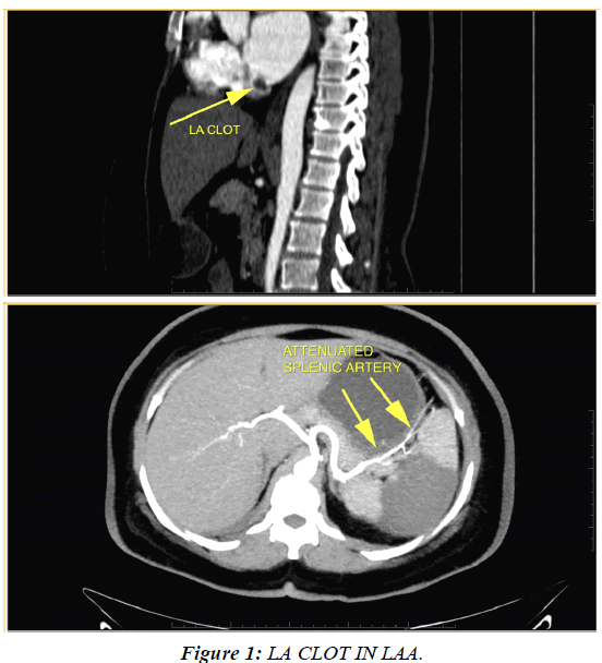
The incidence of LA thrombus in Mitral Stenosis in sinus rhythm is 6.6%. Trans Oesophageal Echo is warranted in MS patients in sinus rhythm when they are more than 44 years. Transthoracic echo is inadequate to diagnose Left Atrial Appendages clot. The left atrial appendage (LAA) is the most common site for LA clot (Figure 1). Left atrial appendage is anatomically attached to the left inferior portion of the left atrium and consists of muscular trabeculae [1,2]. The left atrial appendage does not contribute to a large degree to overall cardiac output. Transesophageal echocardiographic findings of a left atrial appendage thrombus include direct visualization of a mobile echodensity within the appendages. Cardiac CT can be used to diagnose LAA Clots.
Systemic thrombolism in LAA clot is known and can cause stroke, mesentric arterial embolisation or splenic infarction (Figure 2).
Splenic infarction refers to occlusion of the splenic vascular supply, leading to parenchymal ischemia and subsequent tissue necrosis. The infarct may be segmental, or it may be global, involving the entire organ [3]. It is the result of arterial or venous compromise and is associated with a heterogeneous group of diseases (Figure 3) [4].
Discussion
Rheumatic heart disease and Non Rheumatic diseases are the causes of Acute or Chronic Atrial Fibrillation. And it is responsible for thromboembolism. The overall incidence of thromboembolism both systemic and pulmonary is about 30%. Most of the time the transthoracic echo is carried out and clot in atrias are rulled out [5-7].
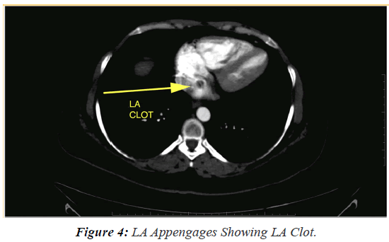
In our case also Tran’s thoracic echo did not show any clots which are seen in LA Appendages (LAA). We carried out Trans esophageal echo which showed clot. Thus the sensitivity of Trans esophageal echo is more than transthoracic echo (Figure 4).
Role of Anticoagulants in both Rheumatic and non-rheumatic atrial fibrillation is proven without doubts. Only antiplatelet therapy is not advocated, which our patient was receiving. New Oral Anticoagulants like Debigatran and Reviroxaben is advocated in Non Valvular Atrial fibrillations. CT scan is another wonderful tool to diagnose atrial clots [8].
All chronic atrial fibrillation patients’ needs anticoagulants in proper dosage along with guidance about food habits, regular Prothrombin time test and dose of anticoagulants are to be decided on INR. Therapeutic level of INR is about 3 to 3.5. Concomitant usages of green leafy vegetables, certain fruits and certain drugs are known to affect the efficacy of anticoagulants [9].
References
- Agmon Y, Bijoy K, Khandheria, et al. Clinical Investigation and Reports. Clinical and Echocardiographic Characteristics of Patients with Left Atrial Thrombus and Sinus Rhythm. 2002;105:27-31.
- SaidiSJ, Motamedi MHK. Incidence and factors influencing left atrial clot in patients with mitral stenosis and normal sinus rhythm. 2004; 90:1342-1343.
- Silaruks S, Thinkhamrop B, Kiatchoosakun S, et al. Dissolving Left Atrial Clots in Patients with Mitral Stenosis. Ann Intern Med. 2004;140:101-105.
- http://www.healio.com/cardiology/learn-the-heart/cardiology-review/topic-reviews/atrial-appendage-thrombus
- Bansal M, Kasliwal R. Mobile Large Left Atrial Thrombus N Engl J Med. 2015; 372:e2
- FukudaS, Watanabe H,Shimada K, et al. Dissolving Left Atrial Clots in Patients withMitral Stenosis journal of cardiology 2011;58:199-314.
- Rana V. Left Atrial Thrombus - A Review 2015
- Kawakami T, Kobayakawa H, Hiroyoshi O, et al. Resolution of left atrial appendage thrombus with apixaban. 2013; 11: 26.
- VenkatesanS. Posts Tagged ‘classification of left atrial clot: ICD 2011; 21.


