Case Report - Otolaryngology Online Journal (2018) Volume 8, Issue 2
Retropharyngeal Metastasis from Thyroid Papillary Carcinoma: Case Report
Pedro Carneiro de Sousa*, Rita Peixoto, Catarina Rato, Pedro Koch, Rosa Saraiva, Nuno Trigueiros-Cunha, Delfim Duarte and Nuno OliveiraDepartment of Otolaryngology, Pedro Hispano Hospital, Unit Health Place of Matosinhos, Portugal
- *Corresponding Author:
- Pedro Carneiro de Sousa
Department of Otolaryngology
Pedro Hispano Hospital
Unit Health Place of Matosinhos
Portugal
Mobile: +351913262235
Email: pedrojmcs@gmail.com
Received date: March 29, 2018; Accepted date: April 07, 2018; Published date: April 16, 2018
Abstract
The presence of a retropharyngeal mass elicits the possibility of a wide range of diagnosis. Inflammatory lesions are the most frequent, but neoplastic causes should also be excluded. Observation: A 56-yearsold man presented with a slow growing left cervical mass. Cervical computed tomography showed cystic and calcified lesions in the left retropharyngeal and supraclavicular spaces. Cytology of the supraclavicular mass revealed papillary thyroid carcinoma cells. Total thyroidectomy with left cervical lymphadenectomy and excision of the retropharyngeal mass were ensued. Histology confirmed retropharyngeal and supraclavicular node metastasis from papillary carcinoma. Discussion: Retropharyngeal node metastization from thyroid papillary carcinoma is very rare, but Otolaryngology should think about this possible diagnosis in the evaluation of a retropharyngeal mass, even in patients already submitted to cervical lymphadenectomy."
Keywords
Thyroid papillary carcinoma; Retropharyngeal lesion; Odynophagia; Node metastasis; Throat
Introduction
Differential diagnosis of a retropharyngeal lesion is very wide. The most frequent etiologies are inflammatory, such as cellulitis, abscess and suppurative lymphadenitis nonetheless, malignant causes may also be found: most often, metastasis from squamous cell carcinomas and non-Hodgkin lymphomas [1].
Metastization of thyroid papillary carcinoma to retropharyngeal lymph nodes is very rare [1,2,3], because it commonly happens to paratracheal and jugular lymph nodes [2,3].
Case Report
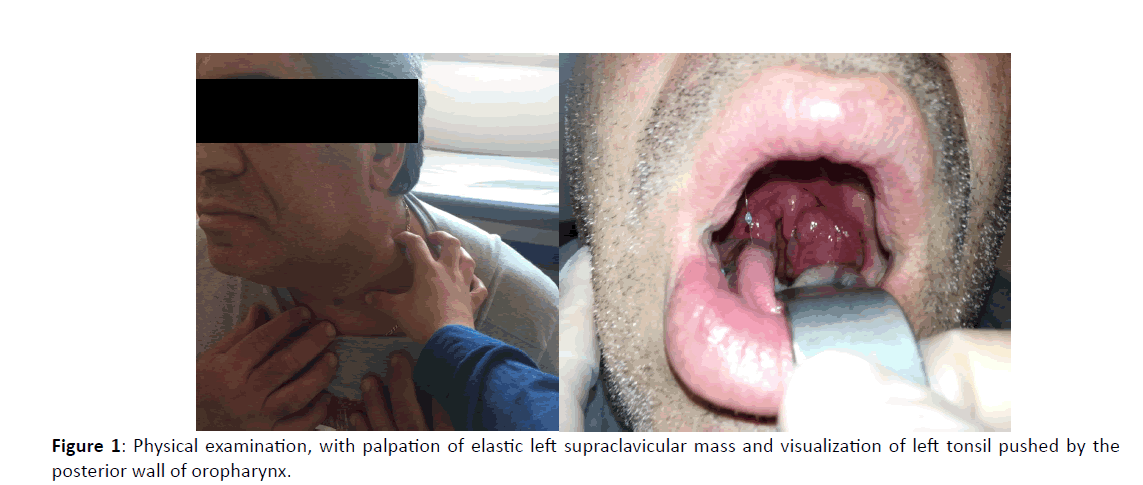
A 56-year-old man presented with a non-painful left cervical mass, with slow growing pattern for more than one year Figure 1. The patient had no complaints of odynophagia, dysphagia, dysphonia and dyspnea. Physical examination revealed a non-dolorous, large, elastic consistency left supraclavicular mass. Oral examination showed a fibrotic and ulcerated left tonsil pushed by the posterior wall of oropharynx Figure 1. A biopsy of the left tonsil was made, but histology revealed only inflammatory features.
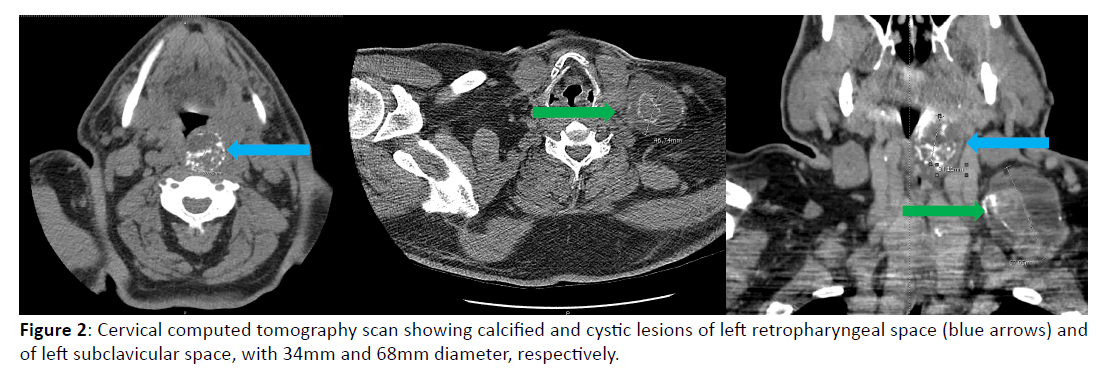
Cervical computed tomography Figure 2 showed a cystic and calcified lesion in the left retropharyngeal space with 34mm, promoting compression of the oropharynx. A bulky 68mm mass with the same characteristics was revealed in the left supraclavicular space as well as a retrosternal goiter with one calcified node.
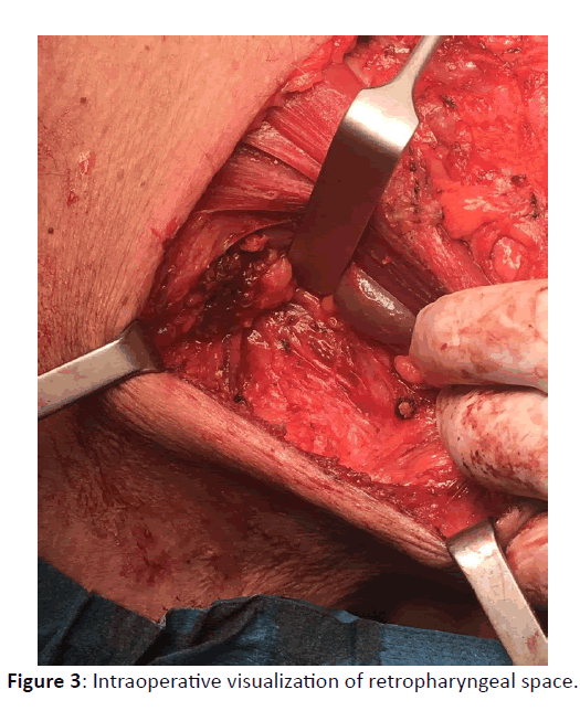
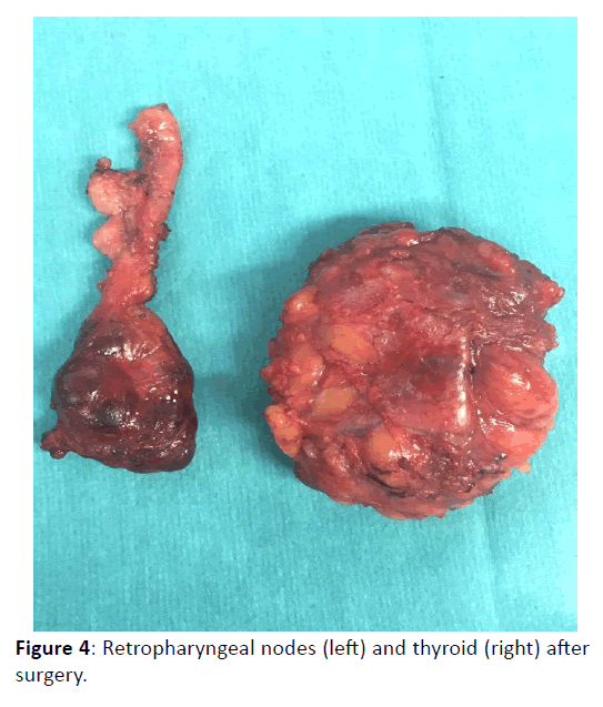
Fine-needle aspirating cytology of the supraclavicular mass revealed papillary thyroid carcinoma cells. The patient was submitted to total thyroidectomy with left cervical lymphadenectomy and excision of the retropharyngeal mass Figures 3 and 4. Both masses noticed in physical examination corresponded to lymph node metastasis of papillary carcinoma.
Discussion
Only 39 cases of retropharyngeal metastasis from thyroid papillary carcinoma with histological confirmation were documented until 2011 [3]. This pattern of metastization results from lymphatic spread: possibly, it comes from retrograde extension from jugular lymph nodes [2,4]. Therefore, it either diagnosis or follow-up after cervical lymphadenectomy, periodic imagiological evaluation (with computed tomography or magnetic resonance imaging) of patients with thyroid papillary carcinoma should be done. In fact, there is a high probability of false negative cases with ultrasonography of neck [4]. The majority of these metastatic retropharyngeal nodes can be resected via transcervical approach, without mandibulotomy [5].
Conclusion
Although retropharyngeal metastasis from thyroid papillary carcinoma are uncommon, Otolaryngology practioners should keep in mind this diagnosis in the work-up of a retropharyngeal mass case. Equally, otolaryngological evaluation of patients unspecified chronic throat symptoms (namely, odynophagia) may allow earlier diagnosis of thyroid papillary carcinoma.
References
- DiLeo MD, Baker KB, Deschler DG, Hayden RE (1998) Metastatic papillary thyroid carcinoma presenting as a retropharyngeal mass. Am J Otolaryngol 19: 404-406.
- McCormack KR, Sheline GE (1970) Retropharyngeal spread of carcinoma of the thyroid. Cancer 26: 1366-1369.
- Kainuma K, Kitoh R, Yoshimura H, Usami S (2011) The first report of bilateral retropharyngeal lymph node metastasis from papillary thyroid carcinoma and review of the literature. Acta Otolaryngol 131: 1341-1348.
- Otsuki N, Nishikawa T, Iwae S, Saito M, Mohri M, et al. (2007) Retropharyngeal node metastasis from papillary thyroid carcinoma. Head Neck 29: 508-511.
- Togashi T, Sugitani I, Toda K, Kawabata K, Takahashi S (2014) Surgical management of retropharyngeal nodes metastases from papillary thyroid carcinoma. World J Surg 38: 2831-2837.


