Mini Review - Journal of Clinical Pathology and Laboratory Medicine (2023) Volume 5, Issue 2
Large Diameter Lipoma of the Large Intestine
Alptekin Sen*Department of Pathology, Istanbul Demiroglu Bilim University Faculty of Medicine, Istanbul, Turkey.
- *Corresponding Author:
- Alptekin Sen
Department of Pathology
Istanbul Demiroglu Bilim University Faculty of Medicine
Istanbul, Turkey.
E-mail: alptekinsen2000@gmail.com
Received: 29-Mar-2023, Manuscript No. AACPLM-23-97294; Editor assigned: 01-Apr-2023, PreQC No. AACPLM-23-97294(PQ); Reviewed: 15-Apr-2023, QC No. AACPLM-23-97294; Revised: 22-Apr-2023, Manuscript No. AACPLM-23-97294(R); Published: 29-Apr-2023, DOI:10.35841/aacplm-5.2.142
Citation: Sen A. Evaluation of different metastasis sites in large intestine adenocarcinoma based on a case. J Clin Path Lab Med. 2023;5(2):142
Abstract
Lipomas although they are benign, non-epithelial, adipose tissue tumors that can be observed throughout the gastrointestinal tract, they are most commonly observed in the colon. Almost all of these lipomas are submucosal, rarely of subserosal or intramucosal origin. In the literature, the incidence of colon lipoma is given as 0.2-4.4%. What makes our case different4 cm, called a 'giant lipoma'. It is above the diameter and the polyp is resected with a part of the large intestine with the preliminary diagnosis.
Keywords
Lipoma, Large intestine, Giant lipoma, Resection.
Introduction
A very important part of colon lipomas is located in the right hemicolon and especially in the cecum. Lipomas that can be observed from a few millimetres to 30 cm in diameter; It is defined as a 'giant lipoma’ on 4 cm.in. This identification may be related to the fact that when they reach a diameter greater than 4 cm [1,2] they cause symptoms these symptoms [3], it may vary from complaints such as abdominal pain and constipation to gastrointestinal bleeding, perforation or obstruction. Our case is a case of large intestine lipoma that was operated with the preliminary diagnosis of polyp but reported as lipoma by us [4].
Case Report
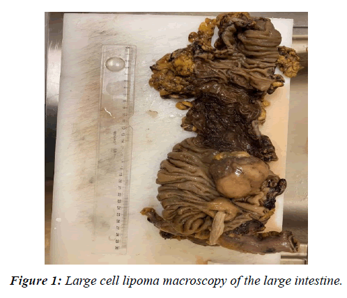
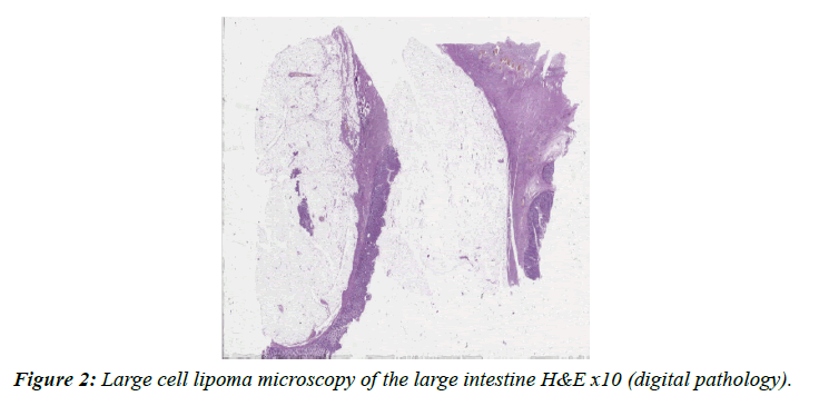
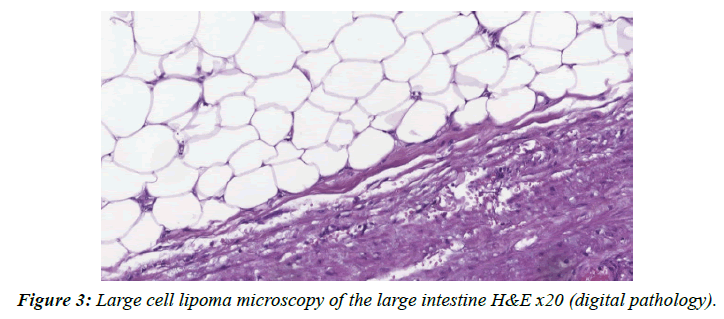
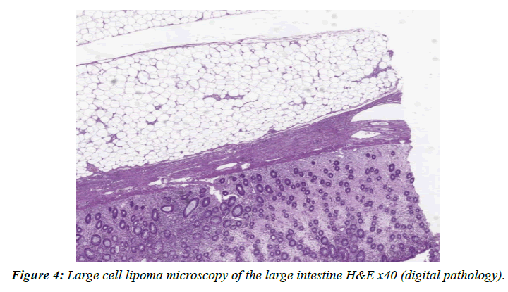
Our case is a 53-year-old male, on July 2022; he was admitted to our General Surgery Clinic with complaints of abdominal pain, abdominal bloating and intermittent longterm constipation that had been going on for about 1 year. As a result of the clinical and radiological evaluation of the patient about 5 cm detected in the transverse colon. Surgical intervention is decided on the mass. The patient is operated on the mass removed with some large intestine was transmitted to our pathology laboratory. The operation material examined in our laboratory is also used to large intestine resection material with a diameter of 30 cm longkta, 1.5 in one surgical end (c1), and 1.5 and in the other surgical end (c2); 1.2 cm in diameter was observed [5]. In the sections of the resection material a fat mass lesion with a diameter of 5 cm. with a diameter of 20 cm from the C1 registered surgical end and 5 cm from the C2 registered surgical end was observed (Figure 1). In areas close to the lesion, in the mucosa of the large intestine, deletion, congestion and atrophy of the ples are observed. In other areas, the intestinal mucosa has a macroscopic normal appearance [6-8]. In the microscopic evaluation, the mass lesion in the forehead bowel resection material described in macroscopy was observed to be a benign natural lipoma structure formed by submucosal located mature fat tissue cells (Figure 2 to Figure 4).
Conclusion and Discussion
Lipomas with adipose tissue tumors, they are rarely detected over 2 cm in diameter and are rarely symptomatic when detected. These symptoms are it can be in the form of abdominal pain, bleeding, diarrhoea or constipation. In GIS although lipoma is not observed very often, when it is observed it is often located in the large intestine. The lipomas of this region rarely reach diameters of 4-5 cm. Although our case is important in this respect, it is also a fact that they should be removed due to their complications such as obstruction, invagination and perforation over 4 cm in diameter. In recent years in the process of removing lipomas. Applications such as 'snare electro surgery' and 'endoloop ligation', which can be applied even if they are 11 cm or larger, have also started to be considered as an option for surgical application. However there is not yet enough information about postoperative recurrence rates in these studies. In addition, the diagnosis made after the procedure once again reveals the importance of pathology. In our case, the preliminary diagnosis was expressed as 'polyp'. In this context, in non-surgical procedures, the suspicion that the diagnosis to be given is 'malignant' forces surgeons to choose the classical operation direction.
Compliance with ethical standards
Appropriate.
Funding
No.
Conflict of Interest
No.
Contributions
There are no contributors other than me.
References
- Vecchio R, Ferrara M, Mosca F, et al. Lipomas of the large bowel. Eur J Surg Oncol. 1996; 162(11):915-9.
- Gordon RT, Beal JM. Lipoma of the colon. Arch Surg 1978; 113(7):897-9.
- Bahadursingh AM, Robbins PL, Longo WE. Giant submucosal sigmoid colon lipoma. Am J Surg. 2003; 186(1):81-2.
- Castro EB, Stearns MW. Lipoma of the large intestine: A review of 45 cases. Dis Colon Rectum. 1972; 15(6):441-4.
- Gould DJ, Morrison CA, Liscum KR, et al. A lipoma of the transverse colon causing intermittent obstruction: a rare cause for surgical intervention. Gastroenterol Hepatol. 2011; 7(7):487.
- Rogy MA, Mirza DF, Kovats E, et al. Pneumatosis cystoides intestinalis (PCI). Int J Colorectal Dis. 1990; 5:120-4.
- Jansen JB, Temmerman A, Tjhie-Wensing JW. Endoscopic removal of large colonic lipomas. Ned Tijdschr Geneeskd. 2010; 154:A2215.
- Waxman I, Saitoh Y, Raju GS, et al. High-frequency probe EUS-assisted endoscopic mucosal resection: A therapeutic strategy for submucosal tumors of the GI tract. Gastrointest Endosc. 2002; 55(1):44-9.
Indexed at, Google Scholar, Cross Ref
Indexed at, Google Scholar, Cross Ref
Indexed at, Google Scholar, Cross Ref
Indexed at, Google Scholar, Cross Ref


