Case Report - Otolaryngology Online Journal (2020) Volume 10, Issue 3
Determination of the Test Retests Reliability of the Otometrics Video Head Impulse Test (Vhit) Among Healthy Adults
Zuraida Z1*, Shamhana AY2, Mohd Normazi Z21Department of Neurosciences, School of Medical Sciences, University Sains Malaysia, Malaysia
2Audiology Programme, School of Health Sciences, University Sains Malaysia, Malaysia
- *Corresponding Author:
- Dr. Zuraida Zainun
Department of Neurosciences
School of Medical Sciences, Health Campus
University Sains Malaysia
16150 Kota Bharu, Kelantan, Malaysia
Tel: +6097673786
Email: drzuraida@yahoo.com (or) zuraidakb@usm.my
Received: May 22, 2020; Accepted: July 20, 2020; Published: July 27, 2020
Abstract
Video head impulse test (vHIT) is a clinical head impulse test to evaluate the vestbulo ocular re?ex (VOR). The VOR is the re?ex mechanism that carries out the functon of gaze stabilizaton which is one of the most important functons of the balance system. Material and Methods: Main aim of this study to determine the test retest reliability of the otometrics video head impulse test (vHIT) among healthy adults aged 18 to 35 years (mean age of 22.51 years). A repeated measures study that recruited 51 young adults (102 ears, 33% were males and 67% were females). All subjects flled Malay Version Vertgo Symptom Scale (MVVSS) as a screening then vHIT testng done for the frst and second session within 1 to 2 weeks. The vHIT was carried out according to the standard protocol and the head of the partcipants were rotated in the di?erent planes which was lateral, anterior and posterior. Results: The lateral canals have high gain compared to anterior and posterior canals in session 1 and session 2. Then, there is no signifcance di?erence (p>0.05 between VOR gain of session 1 and session 2 for each semicircular canals except for the lef lateral. The right lateral showed the highest correlaton ~ 0.746 while right anterior showed the lowest correlaton ~ 0.545. However, the overall canals revealed moderate to high correlaton (0.5 to 0.7). Conclusion: vHIT is a good test to determine the vestbular functon by measuring the VOR gain of each canal.
Keywords
Test retest reliability, Video head impulse test (vhit), Vestibular
Introduction
The video head impulse test (vHIT) is one of tests recently exists in vestibular testing. It is used to identify overt and covert saccades and study gain of vestibulo ocular reflex (VOR) of each semicircular canal (SCCs) separately. In order to diagnose hypo activity of the vestibular labyrinths, clinicians embraced this test as one of the most reliable tests. The head impulse test evaluates the functional status of the VOR. The VOR helps in maintaining gaze on a stationary object while the head is in motion (Bronstein AM and Gresty MA 1991). To describe it in short; it is the biological mechanism that helps to stabilize images in the fovea which is the most sensitive point in the retina. For example, when the visual target moves but the head is steady, the head moves but the visual target is steady or when both the head and the visual target are moving together. During head motion, VOR will control head stability by generating head movements that are equal or opposite to the head motions.
The main components of the VOR consist of semicircular canals, vestibular nuclei and ocular motor nuclei and extra ocular muscles. Utricle, saccule and three SCCs in one side of the head are one part of the peripheral vestibular system. SCCs of both sides of head are connected in orthogonal planes that allows for the detection of head movement in three dimensional axes. The SCCs will detect the angular acceleration and two coplanar canals work together on each side of the head [1]. Left horizontal SCC works in pair with right horizontal SCC and the left anterior (LA) works with right posterior (RP) SCCs or left anterior right posterior (LARP), Biswas [1].
Right anterior (RA) and left posterior (LP) SCCs or right anterior left posterior (RALP) function together while rotation of head in different direction [1]. For example, the discharge rate of the left afferent nerve of horizontal SCC increases when head rotates in the left direction in the horizontal plane and at the same time the discharge rate of the right afferent nerve of horizontal SCC decreases due to the resting discharge rate. The right side of compensatory eye movement of the VOR occurred when the difference in output between the right and left horizontal SCCs happened. So, the eyes will remain still in space during head rotation and enable stable vision.
The video head impulse test is mainly based on two principles of vestibular physiology. They are stimulation of the particular canal that will lead to the eye movements in the plane of same canal and excitatory responses are larger than inhibitory responses. The three SCCs are functionally orthogonal to each other but the head impulse only stimulates one pair of semicircular canal of that particular plane and not the other two pairs of SCCs. VHIT will assess all three pairs of SCCs separately and we can know the exact site of lesion in any three SCCs by measuring VOR gain. VOR gain value of vHIT has been utilized to diagnose different vestibular pathologies like Vestibular Migraine [2], Benign Paroxysmal Positional Vertigo [3] and other diseases. To confidently perform vHIT and interpret the findings of the test, the clinician needs to understand the underlying physiology and the mechanism of the generation of the VOR as well as that of catch-up saccades in patients with poor vestibular function [1]. There are certain basic principles of the neural activity in the vestibular system and how the VOR works. The VOR ensures best vision during head motion by moving the eyes contrary to the head to stabilize the line of sight in space.
The VOR will produce compensatory eye movements that are opposite indirection to head movement to maintain a steady visual image. For example, when the head turns to the right, the eyes move to the left and vice versa. Indeed, head movement in any direction produces an eye movement in the opposite direction [4].
There is a constant neural activity in a subject with normal vestibular function that is generated in the vestibular labyrinths even when the subject is standing erect in a static visual field without moving the head. This constant neural activity is about 100 spikes per second and is known as the resting neural activity of the vestibular labyrinth [1]. So, we know even the head is moved either to the right or left, both the labyrinths are being stimulated. For example if the head is moved to the right side, the vestibular labyrinth on that side is excited and the vestibular labyrinth on the other side will be inhibited. When the head movement is fast enough, then the neural activity in the labyrinth on the side to which the head is moved will increased drastically and the generated neural activity can go up from the resting activity of about 100 spikes per second to 400 or more spikes per second but very soon returns to the resting potential of 100 spikes per second.
The vHIT has a good sensitivity and that is why it is the most recently developed in the world. VHIT requires low amplitude, high velocity head impulse and need cooperation by the patient to wear a pair of lightweight goggles. The vHIT is capable to record overt and covert saccades and the accurate recording will allow clinicians to further review patients’ results. It is sufficiently specific to use in a clinical protocol and it is agreeing with previous research using various vHIT systems [2]. But an adequate training and practice are required to make it more accurate, consistent and comfort with applying the thrusts to a patient. There is significance difference between leftward and rightward thrusts at 60to 80 milliseconds. Specifically, VOR gain for leftward thrusts was greater than gain for rightward thrusts.
In this study, we will determine the validity of VOR gain parameter and peak head velocities in anterior, posterior and lateral vHIT. The test retest reliability of the VOR gain parameters is very important in order to know is it there are any significance differences between VOR gain of session 1 and session 2 for each SCC.
Materials and Methods
The research design for this study is prospective repeated measures by using the purposive sampling was conducted in Otorhinolaryngology Head and Neck Surgery (ORL-HNS) Clinic, Hospital University Sains Malaysia (HUSM), Kubang Kerian, Kelantan.
Study patients those who were no complaint of dizziness and vertigo problems and people with normal balance functions. The respondents were chosen among adults who were 18-35 years old. Between February-May 2019, subjects were recruited and all patients that fulfilled the inclusion criteria were asked to participate in the study after they had signed the written consent form. An ethical approval from Research Ethics Committee (Human) USM was obtained prior to the study.
Malay version of vertigo symptom scale (MVVSS) form
Before choosing the respondents, screening was done by using Malay Version of Vertigo Symptom Scale (MVVSS) by self-administered. They answered all the questions given and after that if they did not have any problems or complaint regarding dizziness or vertigo, the test was preceded for the first session. If their scoring was 14 and above or they complained of dizziness or vertigo, they cannot proceed to the test.
First session of video head impulse test
The first session of the test was done to all the chosen respondents. VHIT was performed using ICS impulse video goggles to record the eye rotation response to an abrupt head rotation in the plane of the canal. Target was kept at the eye-level at a distance of 1m in front of participants. While standing behind of each participant, his/her head was rotated accordingly. Then, the VOR gain for each canal was recorded.
Second session of video head impulse test
VHIT testing was repeated within 1 to 2 weeks to determine the test retest stability of VOR gain between first and second sessions. Eventually, when all the information is gathered and completed from these two sessions, data analysis is performed using MedCalc version 18.11.6 Statistical Software.
Statistical analysis
The statistical analyses performed using MedCalc version 18.11.6 Statistical Software. Both descriptive and inferential statistical analyses were used. Mean and standard deviation were expressed as applicable. The non-parametric method was used to analyze those data as the results were not normally distributed. The responses were recorded for all the participants in two sessions and the responses were present for all the participants in two sessions.
Results
Participants
This research involved 57 participants, aged from 20-31 years old (mean=22.51, SD=1.67) who were eligible and voluntarily participated in present study. Participants were selected specifically and chosen based on the inclusion criteria. Consequently, 6 subjects were excluded due to incomplete results and fail screening of MVVSS. Therefore, the total of participants who were recruited in this study was 51 participants. The mean age of participants was 22.51.
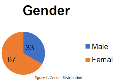
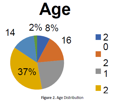
Figure 1 illustrate 67% of the participants were female and the remaining of 33% were male participants (Figure 1). Figure 2 illustrate the age distribution among the participants in the research. Mostly, participants are from age of 23 years old with 37%, followed by 22 years old with 23%, 21 years old with 16%, 24 years old with 14%, 20 years old with 8% and 31 years old with 2% (Figure 2).
In this present study, the results of each participant were analyzed for all six semicircular canals of both ears. Overall, the outcomes of 114 ears were analyzed. However, it was unfortunate that the results of 4 participants were incomplete for second session and had to be excluded while the other two need to be excluded due to fail screening. Therefore, the total of ears that involved in final analysis was 102 ears (51 participants).
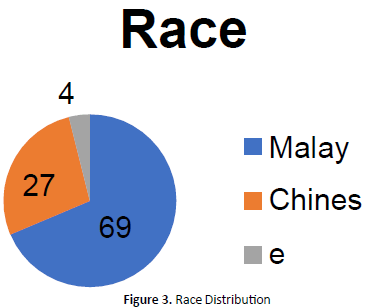
The participants were recruited form Health Campus, University Sains Malaysia, Kubang Kerian, Kelantan. Participants who joined the study consisted of students and staffs from School of Health Sciences such as Dietetics, Speech Pathology, Audiology, Nutrition, Biomedicines and Medical Radiation. There are also some participants from School of Medical Sciences. Figure 3 illustrate the race distribution among the participants in the research. Mostly, participants are Malay with 69%, followed by Chinese with 27% and Indian with 4% (Figure 3).
Interquartile range of VOR gain
Descriptive analysis was done to calculate mean, median, standard deviation and interquartile range of VOR gain in all three planes in both the directions. That is left lateral (LL), right lateral (RL), left anterior (LA), right posterior (RP), right anterior (RA) and left posterior (LP).
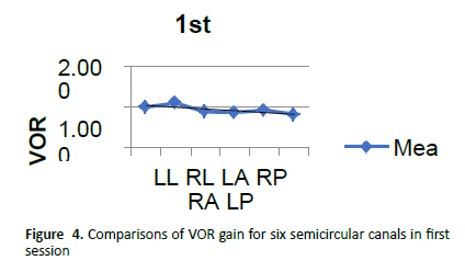
In first session analysis the mean for left lateral is 1.006 with median of 1.000, standard deviation of 0.130 and 0.910 to 1.085 for IQR. For right lateral, the mean is 1.101 with median of 1.075, standard deviation of 0.153 and 1.000 to 1.150 for IQR. While for the left anterior, the mean is 0.879 with median of 0.890, standard deviation of 0.146 and 0.760 to 0.960 for IQR. The mean for right posterior is 0.862 and median is 0.840 with standard deviation of 0.106 and 0.790 to 0.918 for IQR. While the mean for right anterior is 0.925 with median of 0.910, standard deviation of 0.135 and 0.820 to 1.025 for IQR. For the left posterior, the mean is 0.821 while the median is 0.830 with 0.139 of standard deviation and 0.760 to 0.910 of IQR (Table 1).
| Variable | n (%) |
|---|---|
| Age | |
| 20 | 8 |
| 21 | 16 |
| 22 | 23 |
| 23 | 37 |
| 24 | 14 |
| 31 | 2 |
| Gender | |
| Male | 33 |
| Female | 67 |
| Race | |
| Malay | 69 |
| Chinese | 27 |
| Indian | 4 |
Mean=22.51 SD=1.67
Table 1. Demographic data of subjects (n=51)
It can be revealed from Figure 4 that mean gain values for lateral canals are slightly higher compared to the other planes in the first session. However, the mean gain values for the posterior canals are the lowest if compared to the other canals (Figure 4).
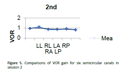
In second session analysis the mean for left lateral is 0.960 with median of 0.940 and standard deviation of 0.132 and 0.870 to 0.998 for IQR. The mean for right lateral is 1.096 with median of 1.075 and standard deviation of 0.140 and 1.000 to 1.150 for IQR. For the left anterior, the mean is 0.858, median is 0.850 with standard deviation is 0.168 and 0.783 to 0.960 for IQR. While the right posterior got 0.861 for the mean with median of 0.860 with standard deviation of 0.118 and 0.793 to 0.918 for IQR. The mean for right anterior is 0.918, median is 0.910 with standard deviation is 0.150 and 0.835 to 0.988 for IQR. For the left posterior, the mean is 0.823 with median of 0.825 and standard deviation of 0.130 and 0.760 to 0.900 for IQR.
Figure 5 illustrated that mean VOR gain for lateral are slightly higher compared to the other planes in the second session. However, the posterior canals are slightly lower if compared to another two planes (Figure 5).
Table 2 showed the p-value and effect size of six semicircular canals between two sessions and wilcoxson signed rank test was administered (Tables 2 & 3). Table 4 showed the p-values and it showed significant difference (p<0.05) for the left lateral (p=0.004). However, the effect size showed 0.349 for the left lateral which is small effect size. As shown, there is no significant difference (p>0.05) for the mean VOR gain value for right lateral (p=0.804) and left anterior (p=0.420) with small effect size 0.043 and 0.131 respectively. Then, there is also no significant difference for the right posterior (p=0.918), right anterior (p=0.870) and left posterior (p=0.526) with small effect size for these canals which are 0.011, 0.050 and 0.072 respectively.
| Canal | LL | RL | LA | RP | RA | LP |
|---|---|---|---|---|---|---|
| p-Value | 0.004 | 0.804 | 0.420 | 0.918 | 0.870 | 0.526 |
| Effect Size | 0.349 | 0.043 | 0.131 | 0.011 | 0.050 | 0.072 |
Table 2. p-value and effect size of six semicircular canals of both sessions
| Canal | LL | RL | LA | RP | RA | LP |
|---|---|---|---|---|---|---|
| r-Value | 0.688 | 0.746 | 0.728 | 0.712 | 0.545 | 0.606 |
| p-Value | P<0.0001 | P<0.0001 | P<0.0001 | P<0.0001 | P<0.0001 | P<0.0001 |
Table 3. r-value and p-value of six semicircular canals of both sessions
| Canal | LL | RL | LA | RP | RA | LP |
|---|---|---|---|---|---|---|
| 90% Range |
0.840 to 1.190 |
0.950 to 1.325 |
0.608 to 1.100 |
0.700 to 1.025 |
0.728 to 1.153 |
0.628 to 0.998 |
Table 4. 90% range of VOR gain for each canals
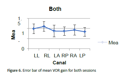
Table 3 showed the r-value and p-value of VOR gain between the first and second session and the Spearman's rank-order correlation was administered. A spearman’s rank order correlation showed statistically significant correlation (p<0.05) in VOR gain for all canals (p<0.0001). As shown in the table 3, there is a high correlation in VOR gain in left lateral (r=0.688), right lateral (r=0.746), left anterior (r=0.728), right posterior (r=0.712), right anterior (r=0.545) and left posterior (r=0.606) between session 1 and session 2.
As illustrated in Figure 6, the right lateral canal showed high positive correlation and the right anterior is the lowest positive correlation. However, overall canals revealed moderate to high correlation as according to the Rule of Thumb [5] (Figure 6). As illustrated in Figure 6, the mean for the VOR gain for lateral is higher compared to the other two canals which are posterior and anterior canals.
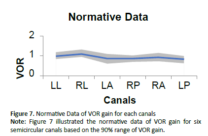
Table 4 showed the 90% range of VOR gain for each canals and the left lateral showed 0.840 to 1.190 ranges while for the right lateral showed 0.950 to 1.325. Then, for the left anterior, the 90% range of VOR gain is 0.608 to 1.1 and for the right posterior is 0.7 to 1.025. For the right anterior, the 90% range is 0.728 to 1.153 and 9.628 to 0.998 for the left posterior (Figure 7).
Discussion
In this current study, the main aim was to determine the test retest reliability of the Otometrics video head impulse test (vHIT). VHIT is a popular test to evaluate the vestibular ocular reflex (VOR). It is also one of the most reliable tests to diagnose hypo activity of the vestibular labyrinths. Therefore, in this study, the VOR gain is intended to determine the test retest reliability of vHIT among healthy adults.
The mean VOR gain for right lateral canal was higher than the left lateral canal, right anterior canal was higher than left anterior canal and right posterior was higher than left posterior canal in present study for session 1 and session 2. McGarvie et al. [6] also reported that the VOR gain for rightwards impulse was slightly higher than leftwards impulses. Similarly, Patterson et al. [7] reported a better VOR gain for the rightward movements compared to the leftward movement using three hand placement techniques. It has been reported that as the head rotates to the right direction, right eye has to make larger rotation comparison to left eye to remain fix at the target by the subjects. Mc Garvie et al. [6] also reported that head movement towards the right causes larger rotation of the right eye and this effect is reversed when head moves towards the left side. Such effects have also been reported by measuring both eyes with dual search coils. This could also be a probable reason for difference in the VOR gain between right and left. Recall that for determining the vHIT results for all semicircular canals in session 1 and session 2 and the specific objective is achieved.
Viirre [8] reported that the magnitude of VOR gain increases with increasing head velocity and decreasing target distance. There could be a probability of difference in the velocity of eye movement in right and left direction which may be causing higher VOR gain in right side impulses for canals stimulation compared to the left. The test also was performed by a right handed and the difference in velocity of head impulses could be because of right handed tester, where right hand dominance would have caused a more velocity of the head impulses towards the right compared to the left. Similar findings of right hand dominance have also been reported by Matiño-Soler [9].
In the present study, gain for the left anterior canal was more compared to the right posterior canal, and this could also be because of the handedness of the clinician wherein the left anterior impulses are easier compared to the right posterior impulses.
From this study among healthy adults in Malaysian populations, the normative values of VOR gain is presented in Table 4 by using 90% range. The VOR gain for lateral canal is between 0.8 to 1.3, but for the vertical canals which are anterior and posterior canals, the normal range is accepted as 0.6 to 1.2 and 0.6 to 1.0 respectively. Based on study from Yollu et al. [10], the normal VOR gain is specified by the manufacturer (ICS optometric vHIT) as > 0.8 for the lateral canals and >0.7 for the vertical canals. If compared to the previous study, the normal range of VOR gain for lateral canal is quite similar to this present study but for the vertical canals, the normal range of VOR gain is started from 0.6 (Table 4).
In this present study, the lateral canals have more gain compared to anterior and posterior canals in session 1 and session 2. Recall that from the specific objective to compare the VOR gain of vHIT results between session 1 and session 2, the VOR gain for lateral canal is higher compared to anterior and posterior canals in sessions 1 and 2. The mean VOR gain for lateral canal, anterior canal and posterior canal range between 0.98-1.09, 0.87-0.90 and 0.83-0.86 respectively, which is almost similar to the study by McGarvie et al. [11], Murnane et al. [12] also reported a larger canal deficits for the vertical planes compared to the lateral planes.
The larger gain observed for the lateral canals may be related to the smaller amplitude of eye movement in the vertical plane than in the lateral plane. In our personal experience also it was always easier to generate head impulses in the lateral direction compared to the vertical planes.
From the Table 2, it showed there is no significance difference between VOR gain of session 1 and session 2 for each semicircular canal except for the left lateral. Even though there is a significance difference for the left lateral, the effect size is small, indicating that there is no genuine difference between two sessions. So, it is still repeatable and it might be happened by chance and because of the data variability. The result of the present study suggests that the gain parameter of vHIT test is reliable. These results supported the study by Murnane et al. [12] which also found that there was no significant difference between the mean canal deficit for session 1 and session 2 for the lateral, anterior, and posterior canals.
From the Table 3, it showed the correlation between each canals between session 1 and session 2 are highly correlated which are the values are near to the 1.0. According to the Rule of Thumb [5] for interpreting the size of a correlation coefficient, the overall values for all six semicircular canals are moderate to high correlated which are in between 0.5 to 0.7. So, we can conclude that the highest the value (near to 1.0), the highest the correlation. However, based on the study from Bansal & Sinha [13], it did not state about the correlation for each semicircular canal between session 1 and session 2. So, in this present study, the correlation for each canals between these two sessions are stated [14-20]. Recall that for determining the correlation of vHIT results between session 1 and session 2 and the specific objective is achieved. Hence, we know that vHIT is relatively new technique in detecting the various vestibular pathologies. In the present study we observed a good reliability of the VOR gain parameter of vHIT test. Thus indicating that the VOR gain functions can be reliably used using vHIT test [21-26].
Conclusion
Video head impulse test (vHIT) is a good test to know the abnormality of the vestibular organ. VHIT can measure the VOR gain in order to diagnose those with hypo activity or vestibular hypo function. It is a quick, non-invasive and extremely reliable test to evaluate the structural and functional integrity of the six semicircular canals in the two vestibular labyrinths.
The present study shows a good reliability of VOR gain functions using vHIT test in normal participants. Taking the results of the present study in to consideration, any significant change in gain parameters would suggest a vestibular pathology and not a simple random variation. This study is believed to be important for other researchers in order to do the future study especially in those with vestibular problems.
References
- Biswas A. Video Head Impulse Test. In: Clinical Audio-Vestibulometry for Otologists and Neurologists (5th Edn). Bhalani Publishing House, Mumbai 2017:405-46.
- MacDougall HG, Weber KP, McGarvie LA, et al. The video head impulse test: diagnostic accuracy in peripheral vestibulopathy. Neurology 2009;73(14):1134-41.
- Blodow A, Heinze M, Bloching MB, et al. Caloric stimulation and video-head impulse testing in Ménière’s disease and vestibular migraine. Acta Otolaryngol 2014;134(12):1239-44.
- Jones SM, Jones TA, Mills KN, et al. Anatomical and Physiological Considerations in Vestibular Dysfunction and Compensation. Seminars in Hearing 2009; 30(4):231-41.
- Mukaka MM. Statistics Corner: A guide to appropriate use of Correlation coefficient in medical research. Malawi Medical Journal 2012;24(3):69-71.
- McGarvie LA, MacDougall HG, Halmagyi GM, et al. The video head impulse test (vHIT) of semicircular canal function–age- dependent normative values of VOR gain in healthy subjects. Front Neurol 2015;8(6):154-60.
- Patterson JN, Bassett AM, Mollak CM, et al. Effects of hand placement technique on the video head impulse test (vHIT) in younger and older adults. OtolNeurotol 2015;36(6):1061-68.
- Viirre E, Tweed D, Milner K, et al. A reexamination of the gain of the vestibuloocular reflex. J Neurophysiol 1986;56(2):439-50.
- Matiño-Soler E, Esteller-More E, Martin-Sanchez JC, et al. Normative data on angular vestibulo-ocular responses in the yaw axis measured using the video head impulse test. OtolNeurotol 2015; 36(3):466-71.
- Yollu U, Uluduz DU, Yilmaz M. Vestibular migraine screening in a migraine- diagnosed patient population, and assessment of vestibulocochlear function. Cl Otolaryngol 2016;42(2):225-33.
- McGarvie LA, Curthoys IS, MacDougall HG, et al. What does the dissociation between the results of video head impulse versus caloric testing reveal about the vestibular dysfunction in Ménière’s disease?. Acta Otolaryngol 2015;135(9):859-65.
- Murnane O, Mabrey H, Pearson A, et al. Normative data and test–retest reliability of the SYNAPSYS video head impulse test. J Am AcadAudiol 2014;25(3):244-52.
- Bansal S, Sinha SK. Assessment of VOR gain function and its test-retest reliability in normal hearing individuals. Eur Arch Otorhinolaryngol 2016;273(10):3167-73.
- Baloh RW, Honrubia V, Konrad HR. Ewald’s second law reevaluated. Acta Otolaryngol 1977; 83(5-6):475-9.
- Böhmer A, Henn V, Suzuki JI. Vestibulo-ocular reflexes after selective plugging of the semicircular canals in the monkey-response plane determinations. Brain Res 1985;326(2):291-8.
- Bronstein AM, Gresty MA. Compensatory eye movements in the presence of conflicting canal and otolith signals. Exp Brain Res 1991;85(3):697-700.
- DeLong AP, Jacobson G. Specificity of the Video Head Impulse Test System (Doctoral dissertation, Vanderbilt University). 2013.
- Estes MS, Blanks RH, Markham CH. Physiologic characteristics of vestibular first-order canal neurons in the cat. I. Response plane determination and resting discharge characteristics. J Neurophysiol 1975;38(5):1232-49.
- Jacobson GP, Neil TS. Balance Function Assessment and Management, Plural Management. 2014.
- Goldberg JM, Fernandez C. Physiology of peripheral neurons innervating semicircular canals of the squirrel monkey. I. Resting discharge and response to constant angular accelerations. J Neurophysiol 1971;34(4):635-60.
- Guerra JG, Pérez FN. Reduction in posterior semicircular canal gain by age in video head impulse testing. Observational study. Acta OtorrinolaringolEsp 2015;6519(15):11-4.
- Martinez-Lopez M, Manrique-Huarte R, Perez-Fernandez N. A puzzle of vestibular physiology in a Meniere’s disease acute attack. Case Rep Otolaryngol 2015;2015:460757.
- Newman-Toker DE, Tehrani ASS, Mantokoudis G, et al. Quantitative video- oculography to help diagnose stroke in acute vertigo and dizziness–toward an ECG for the eyes. Stroke 2013;44:1158-61.
- Redondo-Martínez J, Bécares-Martínez C, Orts-Alborch M, et al. Relationship between video head impulse test (vHIT) and caloric test in patients with vestibular neuritis. Acta OtorrinolaringolEsp 2015;6519(15):142-9.
- Weber KP, Aw ST, Todd MJ, et al. Head impulse test in unilateral vestibular loss: vestibulo-ocular reflex and catch-up saccades. Neurology 2008;70(6):454-63.
- Zhang Y, Chen S, Zhong Z, et al. Preliminary application of video head impulse test in the diagnosis of vertigo. Lin Chung Er Bi Yan Hou Tou Jing Wai Ke Za Zhi 2015;29(12):1053-8.
