Research Article - International Journal of Pure and Applied Zoology (2019) Volume 7, Issue 2
HISTOMORPHOLOGICAL OBSERVATION ON TESTES AND EPIDIDYMIS OF YAK (BOS GRUNNIENS)
- *Corresponding Author:
- Deka A
Department of Anatomy & Histology, College of Veterinary Science, Assam Agricultural University, India
E-mail: dranilvet01@gmail.com
Received 10th May, 2019; Accepted 21st May, 2019; Published 28th May, 2019
DOI: 10.35841/2320-9585.7.9-11
Visit for more related articles at International Journal of Pure and Applied ZoologyAbstract
In present study, one pair of testes and epididymis of Yak (Bos grunniens) was utilized. In cross section of testes, tunica albugenia, tunica vasculosa layer along with cross section of seminiferous tubules were observed. The cross section of seminiferous tubules contained basement membrane, sertoli cells, spermatogonia, spermatids and spermatocytes. The Sertoli cells were pyramidal shaped and rest on basement membrane. The spermatogonia were found among the Sertoli cells. In the cross section of epididymis of pseudo stratified columnar epithelium, stereo cilia, lumen of seminiferous tubules and smooth muscles were observed. The size of the lumen of the seminiferous tubules were larger compared to the size of the seminiferous tubules of testes.
Keywords
Histology; Testes; Epididymis; Yak
Introduction
Yak (Bos grunniens) is a long-haired domesticated animal surviving in the high altitude above 3000 m where other form of agriculture is practically non-existent. They contribute immensely to the socio-economic conditions of the highlanders. Yak provides food, clothing, fuel and the much needed transportation in the hilly terrains. They are the lifeline of the high altitude dwellers. It is originated from the wild Yak (Bos mutus) (Grubb, 2005). The testes are the vital organ of male reproductive system. It has critical role in spermatogenesis. It helps in the preservation of future germ plasm. There are very scanty literatures available worldwide on histomorphological studies of yak testes. Hence the present study was designed to establish a histomorphological observation on the testes of this important animal of high altitude.
Materials and Methods
The presented study was conducted on one pair of testes of Yak (Bos grunniens) of about five to six years old. The testes were obtained from Yak farm, Indian Council of Agricultural Research-National Research Centre on Yak, Dirang, Arunachal Pradesh, India. The testes were collected at the time of castration. After castration, the testes were kept in 10% formalin. Then the tissue samples of testes were processed for paraffin embedding method. The Paraffin sections of five micron thickness were stained with Haematoxylin and Eosin for histomorphological observation (Luna, 1968).
Result and Discussions
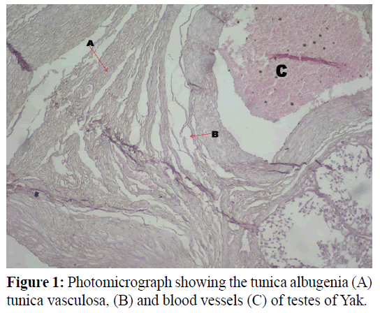
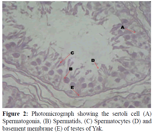
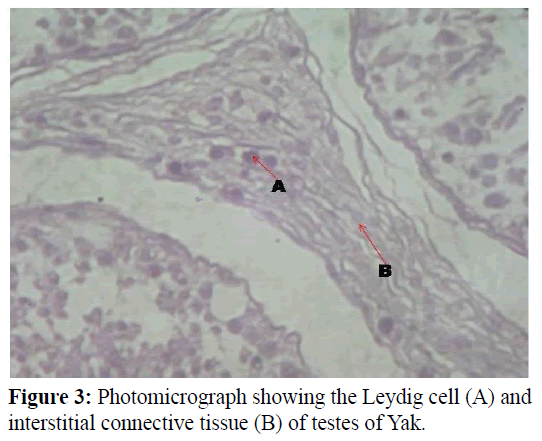
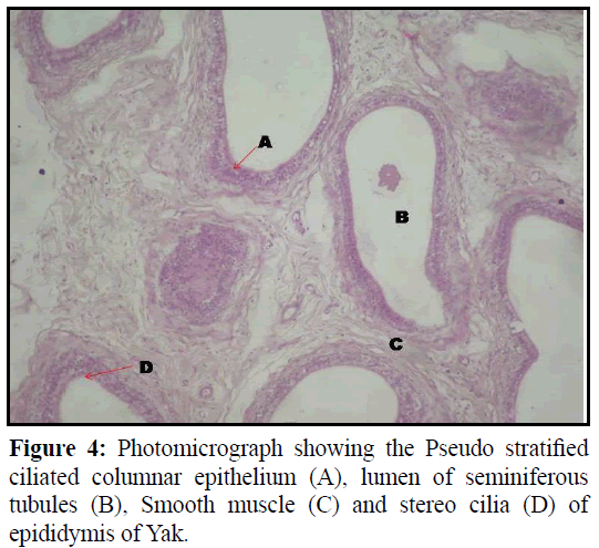
In current studies, one pair of testes and epididymis was utilized. The testes were surrounded by connective tissue layer (Tunica albugenia layer) and blood vessels layer (Tunica vasculosa) (Figure 1). These finding was total agreement with the finding of Moonji and Suwanpugdee (2007) in testis of Rusa deer and Copenhaver (1964) in animal. The tunica albugenia support the testes. Some interstitial connective tissue fibers were observed among the seminiferous tubules along with leydig cells. Some leydig cells were arranged in group and some were arranged in single along those fibers (Figures 2 and 3). These findings were corroborated with the findings of Deka et al., (2014) in Pygmy hog (Porcula salvania) and Elzoghby et al., (2014) in Sheep. Those leydig cells produced testosterone. These hormones give maleness to the animal. The interstitial connective tissue arises formed tunica albugenia. Those connective tissues support the seminiferous tubules. Some blood vessels were observed in the tunica vasculosa layer. Those blood vessels gave nourishment to the testes. The cross section of semiferous tubules contained basement membrane, sertoli cells, spermatogonia, spermatids and spermatocytes (Figure 2). Similarly observation was reported by Gofur et al., (2008) in indigenous bull of Bangladesh and Flore in testes of Human. The sertoli cells had significant role in spermatogenesis, formation of blood testes barrier and inhibit hormone production. Those cells gave nourishment to the spermatogenic cells. Those had were pyramidal shaped and rest on basement membrane. These findings were in accordance with the findings of Mohammed et al., (2011) in testes of indigenous male goat of two years old. The spermatogonia were observed among the sertoli cells because the sertoli cells provide nourishment to the spermatogonia. Similar revealed was reported by Ham (1974) in animal. In the cross section of epididymis of pseudo stratified columnar epithelium, stereo cilia, lumen of seminiferous tubules and smooth muscles were observed (Figure 4). The smooth muscles provide contraction to the seminiferous tubule whereas stereo cilia helped epididymis to ejaculation. The pseudo stratified columnar epithelium, though it was called stratified but it was single layer of cells, actually the shape and size of the cells were irregular and their nuclei were situated at various levels therefore the epithelium appeared to had several layers. The size of the lumen of the seminiferous tubules were larger compared to the size of the seminoferous tubules of testes. Similar result was reported by Semkov et al., (1984) in calves.
Summary and Conclusion
The present study was conducted on the testes and epididymis of Yak (Bos grunniens). The testes were surrounded by Tunica albugenia layer) and Tunica vasculosa. Some interstitial connective tissue fibers were observed among the seminiferous tubules along with leydig cells. Some blood vessels were observed in the tunica vasculosa layer. The cross section of seminiferous tubules contained basement membrane, sertoli cells, spermatogonia, spermatids and spermatocytes. The sertoli cells were pyramidal shaped and rest on basement membrane. The spermatogonia were observed among the sertoli cells. In the cross section of epididymis of pseudo stratified columnar epithelium, stereo cilia, lumen of seminiferous tubules and smooth muscles were observed. The size of the lumen of the seminiferous tubules were larger compared to the size of the seminiferous tubules of testes. These basic observation will be helpful to the scientist, those was engaged in the research of germ plasm preservation of Yak (Bos grunniens).
Acknowledgement
The author is grateful to the Director, ICAR-National Research Centre on Yak, Dirang, Arunachal Pradesh and HOD, Department of Anatomy & Histology, College of Veterinary Science, Assam Agricultural University, Khanapara, Guwahati, Assam, India to carry out the research.
References
- Copenhaver, W. M. (1964). Bailey’s Textbook of Histology. The Williams & Wilkins Company., 15th Edn. 483-515.
- Deka, A., Sarma, K., Das, B. J. (2014). Histomorphological observation on the testes of pygmy hog (Porcula salvania). Indian. Vet. J., 91: 82-83.
- Elzoghby, E. M. A., Sosa, G. A., Mona, N.A.H. (2014). Postnatal development of the sheep testes. Benha Veterinary Medical Journal., 26:186-190.
- Flore, M. S. H. (1986). Atlas of Human Histology. Lea & Febuger Publications, Philadelphia, Maruzen Asia, Singapore, 5th Edn. 208-209.
- Gofur, M. R., Khan, M. Z. I., Karim, M. R., Islam, M.N. (2008). Histomorphology and Histochemistry of testes of indigenous bull (Bos indicus) of Bangladesh. Bangladesh J Veterinary Med., 6:67-74.
- Grubb, P. (2005). "Order Artiodactyla". In Wilson, D.E.; Reeder, D.M (eds.). Mammal Species of the World: A Taxonomic and Geographic Reference. Johns Hopkins University Press., 3rd Edn. 691.
- Ham, A.W. (1974). Histology J.B. Lippincott Company Philadelphia and Toronto., 7th Edn. 900-935.
- Luna, L.G. (1968) Manuals of histological staining methods of Armed forces institute of pathology, 3rd Edn. Mc Graw Hill Book Co., London.
- Mohammed, A. H. S., Kadium, D. A. H., Ebed, A.K. (2011). Some morphometric and histological description of the seminiferous, striaghted and rete testes tubules in the testes of indigenous male goats (two years old). Kufa J for Veterinary Medical Science., 2: 19-29.
- Moonji, P., Suwanpugdee, A. (2007). Histological structure of testis and ductus epididymis of Rusa deer (Cervus timorrensis). Kasetsart Journal., 41: 86-90.
- Semkov, M., Kovachev, K., Zhuroval, D. (1984). Histological characteristics of the testis and epididymis of calves during postnatal development. Vet. Med. Nauki., 21: 76-80.


