Editorial - Journal of Gastroenterology and Digestive Diseases (2016) Volume 1, Issue 1
Pocket-Size Ultrasound: Stethoscope of 21st Century.
- *Corresponding Author:
- Naganuma H
Department of Gastroenterology, Yokote Municipal Hospital, Japan
E-mail: hiron@vesta.ocn.ne.jp
Received date: April 18, 2016; Accepted date: April 26, 2016; Published date: April 29, 2016
Abstract
Ultrasound (US) is the first diagnostic tool use for observing the whole abdomen, and we know that many abdominal diseases have been immediately detected by abdominal US. However, the actual US machine usually weights more than 100 kg and measures 1 by 1.5 meters. It prevents us to bring the machine to bedside anytime and anywhere.
Introduction
Ultrasound (US) is the first diagnostic tool use for observing the whole abdomen, and we know that many abdominal diseases have been immediately detected by abdominal US. However, the actual US machine usually weights more than 100 kg and measures 1 by 1.5 meters. It prevents us to bring the machine to bedside anytime and anywhere.
The development in US technology has recently introduced pocket US, and its diagnostic contribution has been supported by cardiologists who decide therapeutic decision based on findings provided by pocket US (Kitada, Fukuda, & Watanabe, 2013). On the other hand, there is a marked paucity of contribution of pocket US in abdominal field in the literature. This editorial discusses some possible roles of this new diagnostic tool in abdominal field.
Emergency
Pocket US is expected to facilitate the prompt diagnosis of acute abdomen. Use of pocket US is especially recommended for critically ill patients whom we cannot move immediately to the examination room. In this situation, other imagings are not available, only pocket US can be used instead (Mariani & Setla, 2011). In connection with some other available information (history, physical examination, and if possible laboratory data), we will immediately suspect or rule out the usual causes of the symptom.
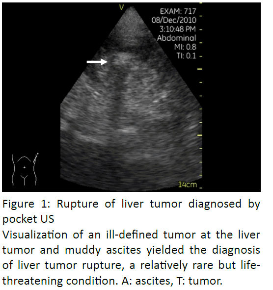
Abdominal US by conventional machine has shown a very high (more than 80-90%) sensitivity and specificity for diagnosing acute abdomen, such as acute cholecystitis, abdominal aorta rupture and liver tumor rupture (Figure 1).
Let us emphasize that delayed management and mortality and morbidity are linked in ill patients. In critically ill, where pocket US will prove contributive.
Outpatient clinic
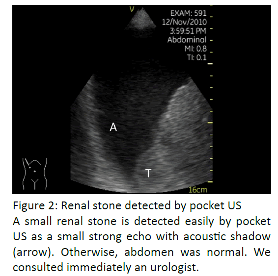
As is in other applications, pocket US yields an excellent clinical decision-making. Abdominal symptoms (discomfort, pain, etc.) are very common in outpatients. Presentation may differ from patient to patient. The physical examination can be misleadingly non-problematic, even with very severe conditions, especially in older patients. A broader differential diagnosis must be considered in patients with abdominal symptom. The diagnosis includes not only common abdominal diseases but also urinary tract problems (stone, urinary retention, etc.), or gynecological diseases (Ovarial torsion, etc.). Recently, its diagnostic contribution has been supported by gastroenterologists who decide next approach based on findings provided by pocket US. According to their preliminary report, about 20% of patients who visit hospital with abdominal symptom are due to urogenital or gynecological diseases (Figure 2) (Ishida, Komatsuda, & Furukawa, 2014).
Renal stone detected by pocket US. A small renal stone is detected easily by pocket US as a small strong echo with acoustic shadow (arrow). Otherwise, abdomen was normal. We consulted immediately an urologist.
Home medical care
In this situation, we face aged patients. They feel difficulties to express themselves. The risk would be to judge the “true” severity based on the patient’s physical examination. Aging may alter the patient’s physical reaction, making evaluation of their true condition more difficult. However, a short delay in detection of diseases may worsen the outcome. Pocket US is the only medical imaging available, and without US information, the clinicians tend to provide a therapy according to standard recommendations. However, pocket US serves to detect incidentally some important findings, such as malignant tumor, liver cirrhosis, lymph node swelling, ascites, and pleural effusion. Detection of such findings leads to an appropriate patient care.
Discussion
Traditionally, clinicians’ physical examinations of the abdomen consist of inspection, auscultation, percussion and palpation in a daily practice. But those are all indirect and on may question whether these traditional methods are sufficiently for examining patients with abdominal disease in 21st century. Until recently, we had no scientific eye in hand, but at present, we have gained it. Pocket US opens a new era to patient care in daily practice. The most important question is “How to use it”.
Conclusion
Pocket US is expected to yield a rapid and accurate diagnostic evaluation in abdominal field, but use of it for abdominal exploration is still not widespread. Basic training in abdominal ultrasound is indispensable, and a systemic training system is important to develop integration of pocket US in daily practice (Andersen, Graven, & Skjetne, 2015).
References
- Kitada, R., Fukuda, S., &Watanabe, H.(2013)Diagnostic accuracy and cost-effectiveness of a pocket-sized transthoracic echocardiographic imaging device. Clin Cardiol36, 603-610.
- Mariani, P.J., &Setla, J.A., (2011) Palliative ultrasound for home care hospice patients. Acad Emerg Med, 2010, 17, 293-296.
- Ishida, H., Komatsuda, T., &Furukawa, K.(2014) Pocket-sized US in outpatient?s clinics. J Med Ultrason (suppl) 38, S402.
- Andersen, G.N., Graven, T., &Skjetne, K., (2015) Diagnostic influence of routine point-of-care pocket-size ultrasound examinations performed by medical residents. J Ultrasound Med34,627-636

